Platinum »
PDB 1a2e-1sj7 »
1eu8 »
Platinum in PDB 1eu8: Structure of Trehalose Maltose Binding Protein From Thermococcus Litoralis
Protein crystallography data
The structure of Structure of Trehalose Maltose Binding Protein From Thermococcus Litoralis, PDB code: 1eu8
was solved by
J.Diez,
K.Diederichs,
G.Greller,
R.Horlacher,
W.Boos,
W.Welte,
with X-Ray Crystallography technique. A brief refinement statistics is given in the table below:
| Resolution Low / High (Å) | 20.00 / 1.90 |
| Space group | P 21 21 21 |
| Cell size a, b, c (Å), α, β, γ (°) | 59.243, 81.526, 86.455, 90.00, 90.00, 90.00 |
| R / Rfree (%) | 19.8 / 23.2 |
Other elements in 1eu8:
The structure of Structure of Trehalose Maltose Binding Protein From Thermococcus Litoralis also contains other interesting chemical elements:
| Chlorine | (Cl) | 5 atoms |
Platinum Binding Sites:
The binding sites of Platinum atom in the Structure of Trehalose Maltose Binding Protein From Thermococcus Litoralis
(pdb code 1eu8). This binding sites where shown within
5.0 Angstroms radius around Platinum atom.
In total 3 binding sites of Platinum where determined in the Structure of Trehalose Maltose Binding Protein From Thermococcus Litoralis, PDB code: 1eu8:
Jump to Platinum binding site number: 1; 2; 3;
In total 3 binding sites of Platinum where determined in the Structure of Trehalose Maltose Binding Protein From Thermococcus Litoralis, PDB code: 1eu8:
Jump to Platinum binding site number: 1; 2; 3;
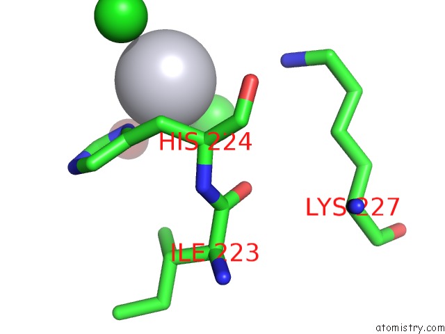
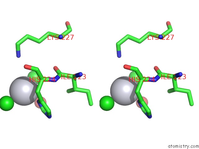
Platinum binding site 1 out of 3 in 1eu8
Go back to
Platinum binding site 1 out
of 3 in the Structure of Trehalose Maltose Binding Protein From Thermococcus Litoralis
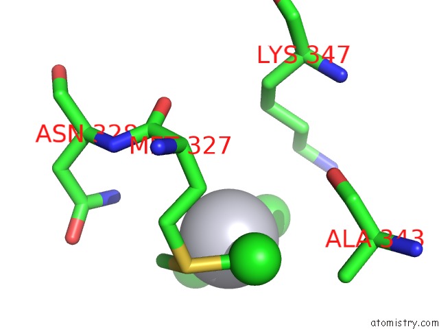
Mono view
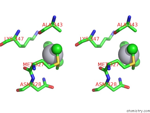
Stereo pair view
Mono view
Stereo pair view
A full contact list of Platinum with other atoms in the Pt binding
site number 1 of Structure of Trehalose Maltose Binding Protein From Thermococcus Litoralis within 5.0Å range:
|
Platinum binding site 2 out of 3 in 1eu8
Go back to
Platinum binding site 2 out
of 3 in the Structure of Trehalose Maltose Binding Protein From Thermococcus Litoralis
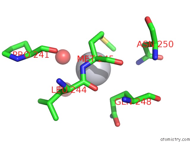
Mono view
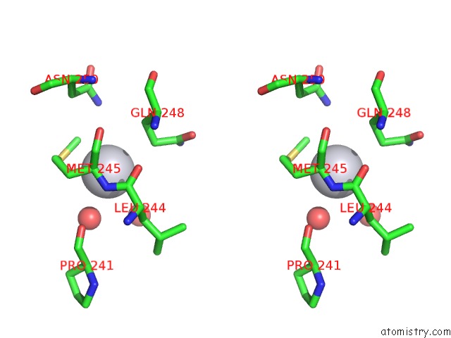
Stereo pair view
Mono view
Stereo pair view
A full contact list of Platinum with other atoms in the Pt binding
site number 2 of Structure of Trehalose Maltose Binding Protein From Thermococcus Litoralis within 5.0Å range:
|
Platinum binding site 3 out of 3 in 1eu8
Go back to
Platinum binding site 3 out
of 3 in the Structure of Trehalose Maltose Binding Protein From Thermococcus Litoralis
Mono view
Stereo pair view
Mono view
Stereo pair view
A full contact list of Platinum with other atoms in the Pt binding
site number 3 of Structure of Trehalose Maltose Binding Protein From Thermococcus Litoralis within 5.0Å range:
|
Reference:
J.Diez,
K.Diederichs,
G.Greller,
R.Horlacher,
W.Boos,
W.Welte.
The Crystal Structure of A Liganded Trehalose/Maltose-Binding Protein From the Hyperthermophilic Archaeon Thermococcus Litoralis at 1.85 A. J.Mol.Biol. V. 305 905 2001.
ISSN: ISSN 0022-2836
PubMed: 11162101
DOI: 10.1006/JMBI.2000.4203
Page generated: Thu Oct 10 10:34:41 2024
ISSN: ISSN 0022-2836
PubMed: 11162101
DOI: 10.1006/JMBI.2000.4203
Last articles
Fe in 2YXOFe in 2YRS
Fe in 2YXC
Fe in 2YNM
Fe in 2YVJ
Fe in 2YP1
Fe in 2YU2
Fe in 2YU1
Fe in 2YQB
Fe in 2YOO