Platinum »
PDB 1a2e-1sj7 »
1rr7 »
Platinum in PDB 1rr7: Crystal Structure of the Middle Operon Regulator Protein of Bacteriophage Mu
Protein crystallography data
The structure of Crystal Structure of the Middle Operon Regulator Protein of Bacteriophage Mu, PDB code: 1rr7
was solved by
M.Kumaraswami,
M.M.Howe,
H.W.Park,
with X-Ray Crystallography technique. A brief refinement statistics is given in the table below:
| Resolution Low / High (Å) | 30.00 / 2.20 |
| Space group | P 32 2 1 |
| Cell size a, b, c (Å), α, β, γ (°) | 81.631, 81.631, 44.843, 90.00, 90.00, 120.00 |
| R / Rfree (%) | 25.2 / 26.8 |
Platinum Binding Sites:
The binding sites of Platinum atom in the Crystal Structure of the Middle Operon Regulator Protein of Bacteriophage Mu
(pdb code 1rr7). This binding sites where shown within
5.0 Angstroms radius around Platinum atom.
In total 2 binding sites of Platinum where determined in the Crystal Structure of the Middle Operon Regulator Protein of Bacteriophage Mu, PDB code: 1rr7:
Jump to Platinum binding site number: 1; 2;
In total 2 binding sites of Platinum where determined in the Crystal Structure of the Middle Operon Regulator Protein of Bacteriophage Mu, PDB code: 1rr7:
Jump to Platinum binding site number: 1; 2;
Platinum binding site 1 out of 2 in 1rr7
Go back to
Platinum binding site 1 out
of 2 in the Crystal Structure of the Middle Operon Regulator Protein of Bacteriophage Mu
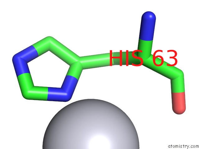
Mono view
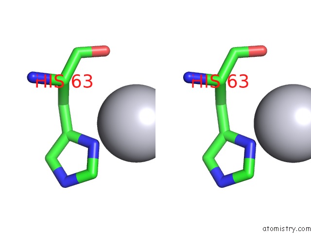
Stereo pair view
Mono view
Stereo pair view
A full contact list of Platinum with other atoms in the Pt binding
site number 1 of Crystal Structure of the Middle Operon Regulator Protein of Bacteriophage Mu within 5.0Å range:
|
Platinum binding site 2 out of 2 in 1rr7
Go back to
Platinum binding site 2 out
of 2 in the Crystal Structure of the Middle Operon Regulator Protein of Bacteriophage Mu
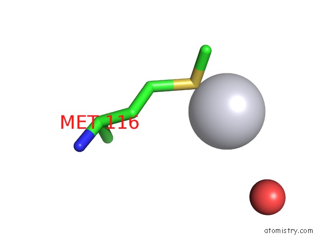
Mono view
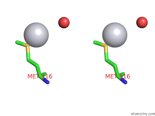
Stereo pair view
Mono view
Stereo pair view
A full contact list of Platinum with other atoms in the Pt binding
site number 2 of Crystal Structure of the Middle Operon Regulator Protein of Bacteriophage Mu within 5.0Å range:
|
Reference:
M.Kumaraswami,
M.M.Howe,
H.W.Park.
Crystal Structure of the Mor Protein of Bacteriophage Mu, A Member of the Mor/C Family of Transcription Activators. J.Biol.Chem. V. 279 16581 2004.
ISSN: ISSN 0021-9258
PubMed: 14729670
DOI: 10.1074/JBC.M313555200
Page generated: Thu Oct 10 10:37:53 2024
ISSN: ISSN 0021-9258
PubMed: 14729670
DOI: 10.1074/JBC.M313555200
Last articles
Cl in 8EFZCl in 8EDD
Cl in 8EDE
Cl in 8EFJ
Cl in 8EBK
Cl in 8EA5
Cl in 8E9Y
Cl in 8EAD
Cl in 8EB3
Cl in 8E6A