Platinum »
PDB 1uas-3co3 »
2b7k »
Platinum in PDB 2b7k: Crystal Structure of Yeast SCO1
Protein crystallography data
The structure of Crystal Structure of Yeast SCO1, PDB code: 2b7k
was solved by
C.Abajian,
A.C.Rosenzweig,
with X-Ray Crystallography technique. A brief refinement statistics is given in the table below:
| Resolution Low / High (Å) | 32.62 / 1.80 |
| Space group | P 1 21 1 |
| Cell size a, b, c (Å), α, β, γ (°) | 54.700, 81.300, 79.500, 90.00, 90.00, 90.00 |
| R / Rfree (%) | 22 / 23.8 |
Platinum Binding Sites:
The binding sites of Platinum atom in the Crystal Structure of Yeast SCO1
(pdb code 2b7k). This binding sites where shown within
5.0 Angstroms radius around Platinum atom.
In total 2 binding sites of Platinum where determined in the Crystal Structure of Yeast SCO1, PDB code: 2b7k:
Jump to Platinum binding site number: 1; 2;
In total 2 binding sites of Platinum where determined in the Crystal Structure of Yeast SCO1, PDB code: 2b7k:
Jump to Platinum binding site number: 1; 2;
Platinum binding site 1 out of 2 in 2b7k
Go back to
Platinum binding site 1 out
of 2 in the Crystal Structure of Yeast SCO1
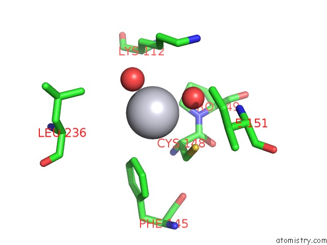
Mono view
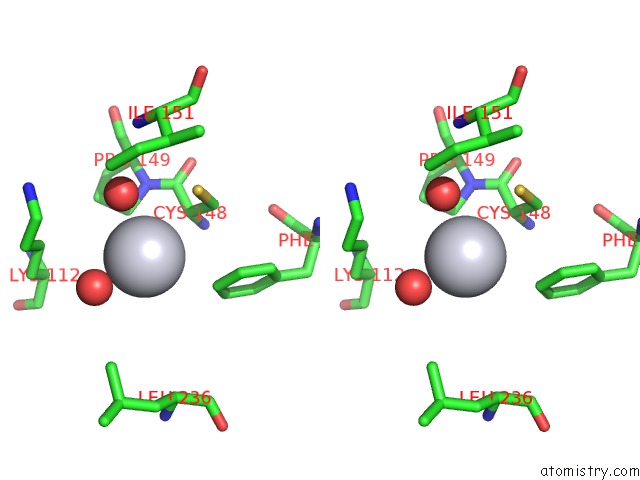
Stereo pair view
Mono view
Stereo pair view
A full contact list of Platinum with other atoms in the Pt binding
site number 1 of Crystal Structure of Yeast SCO1 within 5.0Å range:
|
Platinum binding site 2 out of 2 in 2b7k
Go back to
Platinum binding site 2 out
of 2 in the Crystal Structure of Yeast SCO1
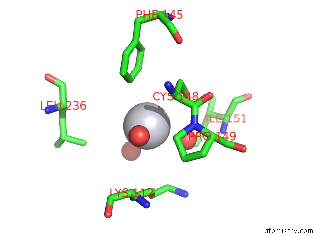
Mono view
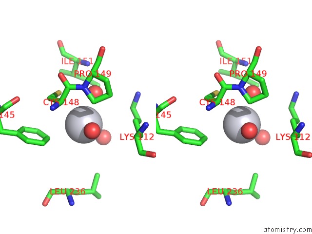
Stereo pair view
Mono view
Stereo pair view
A full contact list of Platinum with other atoms in the Pt binding
site number 2 of Crystal Structure of Yeast SCO1 within 5.0Å range:
|
Reference:
C.Abajian,
A.C.Rosenzweig.
Crystal Structure of Yeast SCO1. J.Biol.Inorg.Chem. V. 11 459 2006.
ISSN: ISSN 0949-8257
PubMed: 16570183
DOI: 10.1007/S00775-006-0096-7
Page generated: Thu Oct 10 10:41:27 2024
ISSN: ISSN 0949-8257
PubMed: 16570183
DOI: 10.1007/S00775-006-0096-7
Last articles
Cl in 2WTUCl in 2WQL
Cl in 2WTA
Cl in 2WSM
Cl in 2WT8
Cl in 2WSL
Cl in 2WSA
Cl in 2WSJ
Cl in 2WS7
Cl in 2WS6