Platinum »
PDB 1uas-3co3 »
2fyf »
Platinum in PDB 2fyf: Structure of A Putative Phosphoserine Aminotransferase From Mycobacterium Tuberculosis
Enzymatic activity of Structure of A Putative Phosphoserine Aminotransferase From Mycobacterium Tuberculosis
All present enzymatic activity of Structure of A Putative Phosphoserine Aminotransferase From Mycobacterium Tuberculosis:
2.6.1.52;
2.6.1.52;
Protein crystallography data
The structure of Structure of A Putative Phosphoserine Aminotransferase From Mycobacterium Tuberculosis, PDB code: 2fyf
was solved by
F.Coulibaly,
E.Lassalle,
E.N.Baker,
Mycobacterium Tuberculosisstructural Proteomics Project (Xmtb),
with X-Ray Crystallography technique. A brief refinement statistics is given in the table below:
| Resolution Low / High (Å) | 19.93 / 1.50 |
| Space group | P 21 21 21 |
| Cell size a, b, c (Å), α, β, γ (°) | 77.478, 94.056, 101.063, 90.00, 90.00, 90.00 |
| R / Rfree (%) | 14.4 / 16.6 |
Other elements in 2fyf:
The structure of Structure of A Putative Phosphoserine Aminotransferase From Mycobacterium Tuberculosis also contains other interesting chemical elements:
| Chlorine | (Cl) | 9 atoms |
Platinum Binding Sites:
The binding sites of Platinum atom in the Structure of A Putative Phosphoserine Aminotransferase From Mycobacterium Tuberculosis
(pdb code 2fyf). This binding sites where shown within
5.0 Angstroms radius around Platinum atom.
In total 6 binding sites of Platinum where determined in the Structure of A Putative Phosphoserine Aminotransferase From Mycobacterium Tuberculosis, PDB code: 2fyf:
Jump to Platinum binding site number: 1; 2; 3; 4; 5; 6;
In total 6 binding sites of Platinum where determined in the Structure of A Putative Phosphoserine Aminotransferase From Mycobacterium Tuberculosis, PDB code: 2fyf:
Jump to Platinum binding site number: 1; 2; 3; 4; 5; 6;
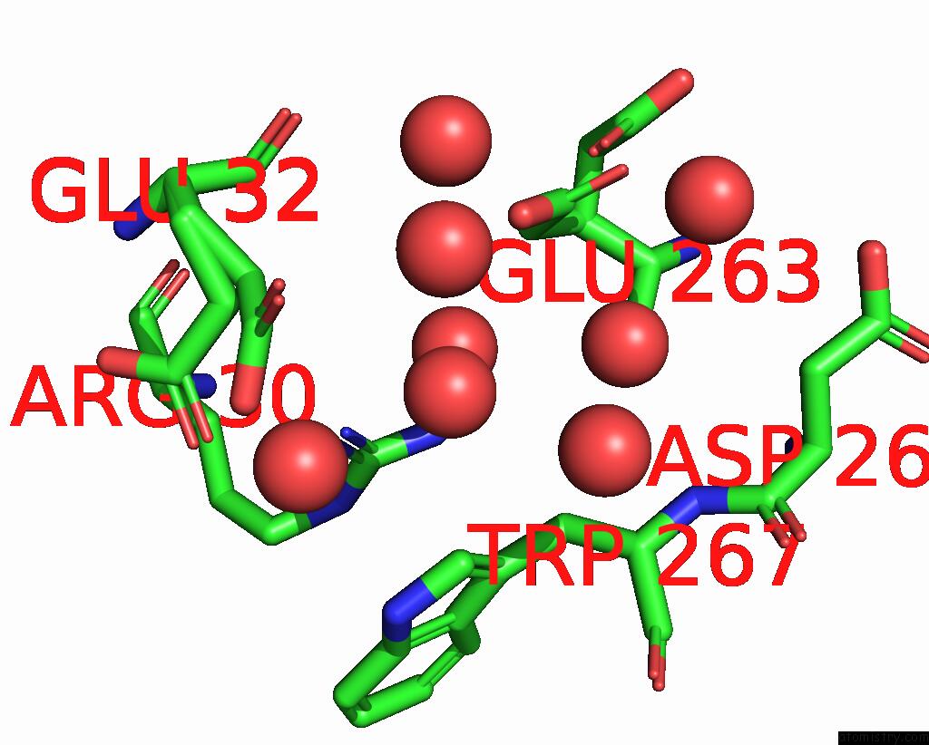
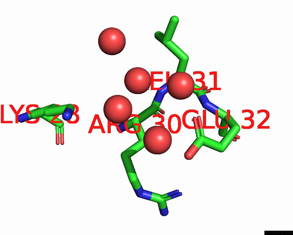
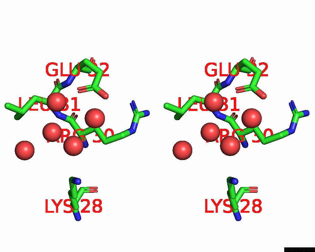
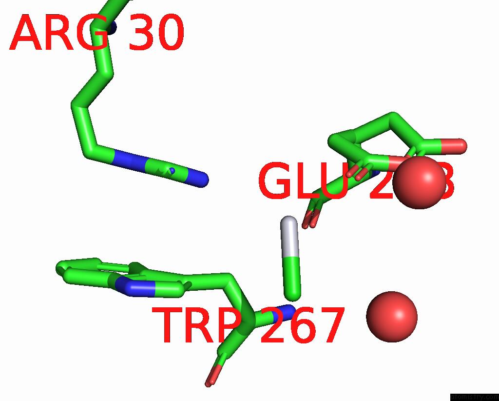
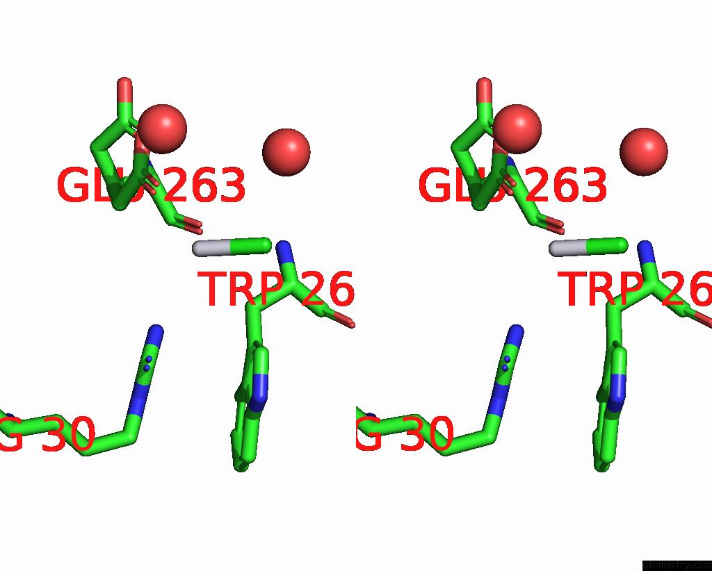
Platinum binding site 1 out of 6 in 2fyf
Go back to
Platinum binding site 1 out
of 6 in the Structure of A Putative Phosphoserine Aminotransferase From Mycobacterium Tuberculosis
Mono view
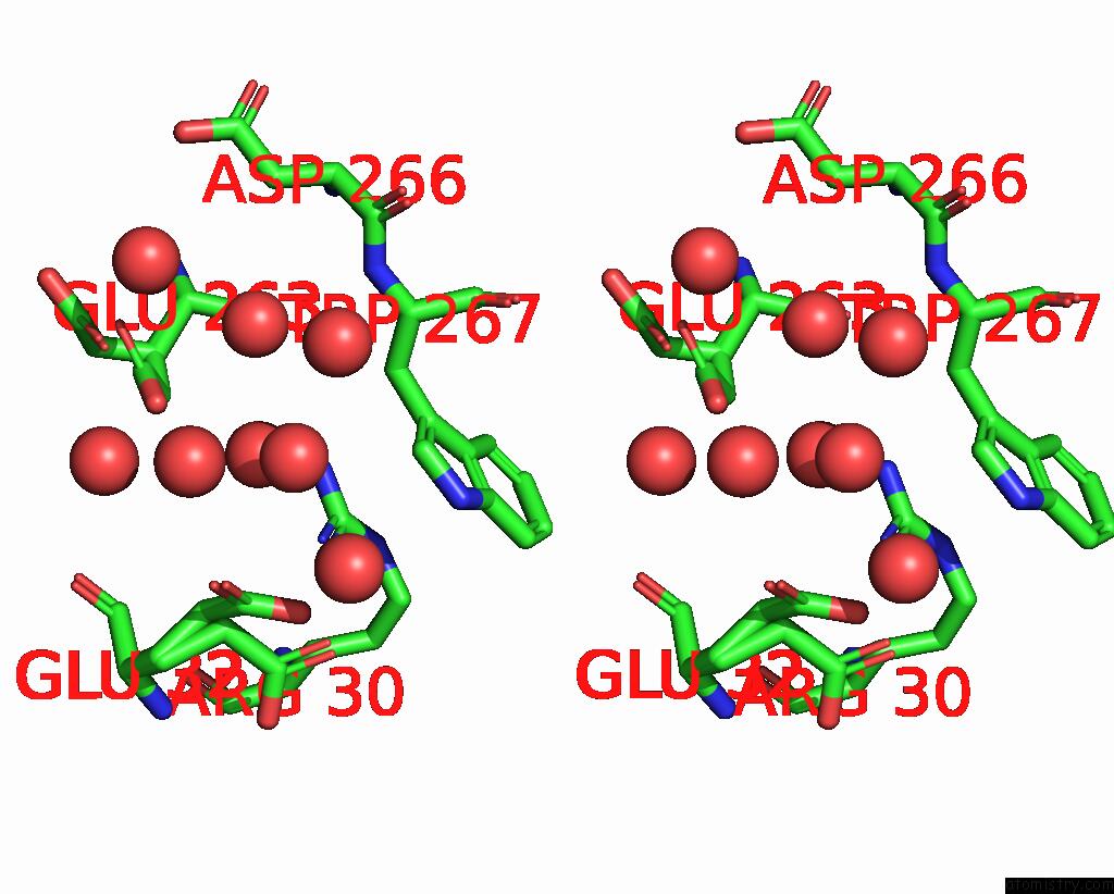
Stereo pair view
Mono view
Stereo pair view
A full contact list of Platinum with other atoms in the Pt binding
site number 1 of Structure of A Putative Phosphoserine Aminotransferase From Mycobacterium Tuberculosis within 5.0Å range:
|
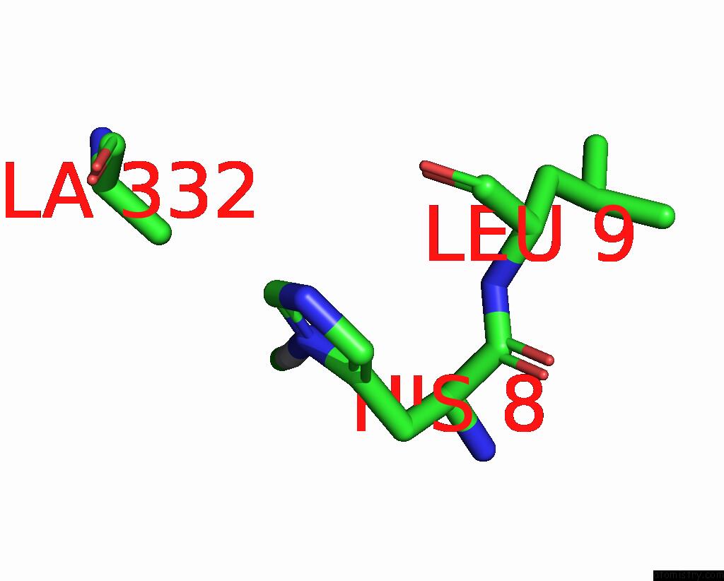
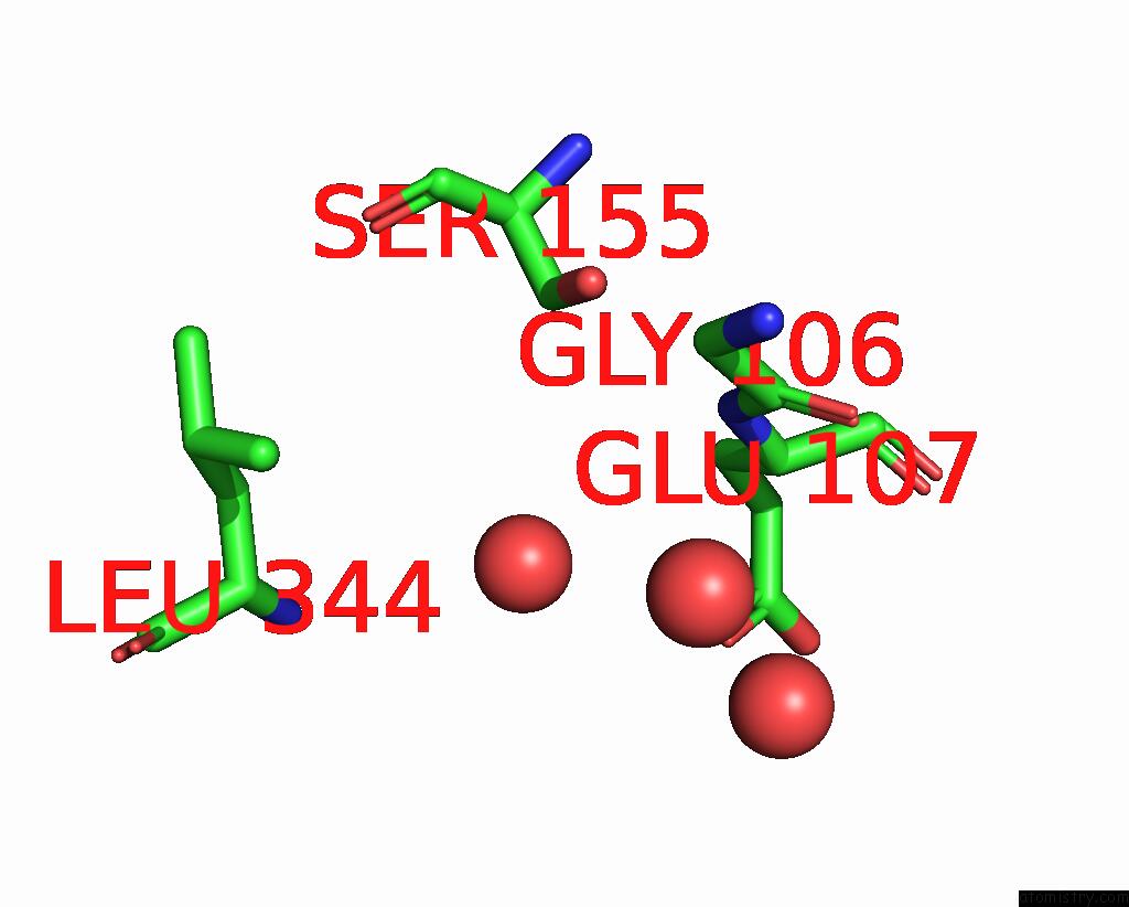
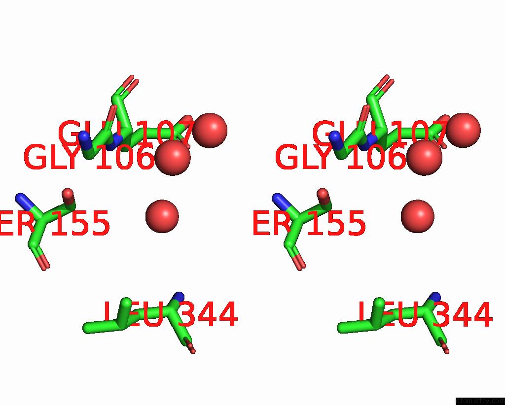
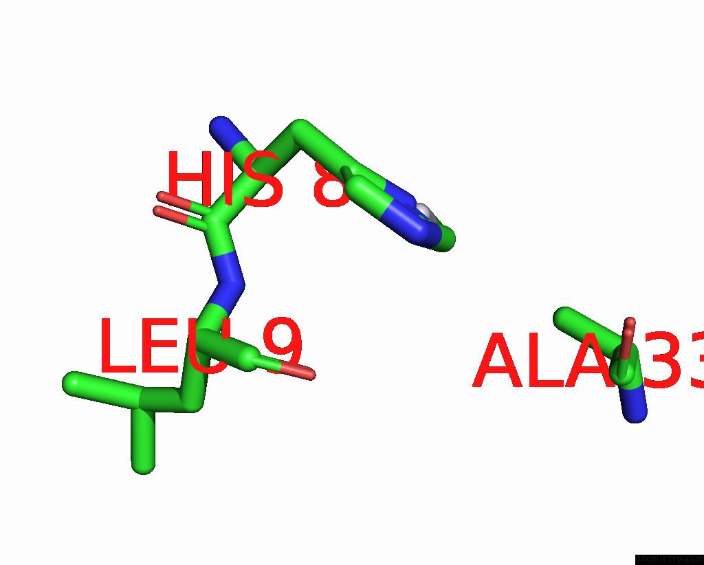
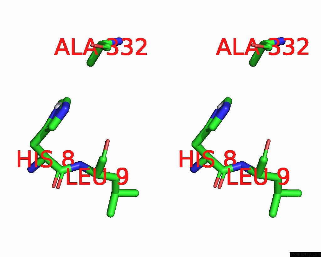
Platinum binding site 2 out of 6 in 2fyf
Go back to
Platinum binding site 2 out
of 6 in the Structure of A Putative Phosphoserine Aminotransferase From Mycobacterium Tuberculosis
Mono view
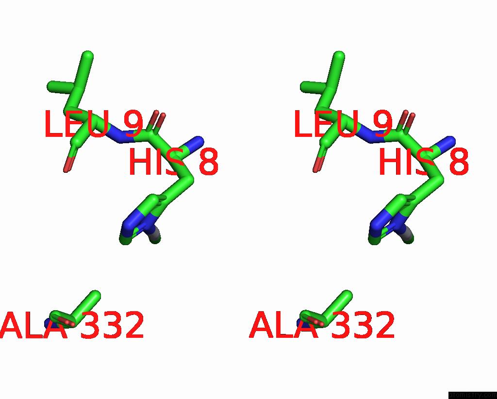
Stereo pair view
Mono view
Stereo pair view
A full contact list of Platinum with other atoms in the Pt binding
site number 2 of Structure of A Putative Phosphoserine Aminotransferase From Mycobacterium Tuberculosis within 5.0Å range:
|
Platinum binding site 3 out of 6 in 2fyf
Go back to
Platinum binding site 3 out
of 6 in the Structure of A Putative Phosphoserine Aminotransferase From Mycobacterium Tuberculosis
Mono view
Stereo pair view
Mono view
Stereo pair view
A full contact list of Platinum with other atoms in the Pt binding
site number 3 of Structure of A Putative Phosphoserine Aminotransferase From Mycobacterium Tuberculosis within 5.0Å range:
|
Platinum binding site 4 out of 6 in 2fyf
Go back to
Platinum binding site 4 out
of 6 in the Structure of A Putative Phosphoserine Aminotransferase From Mycobacterium Tuberculosis
Mono view
Stereo pair view
Mono view
Stereo pair view
A full contact list of Platinum with other atoms in the Pt binding
site number 4 of Structure of A Putative Phosphoserine Aminotransferase From Mycobacterium Tuberculosis within 5.0Å range:
|
Platinum binding site 5 out of 6 in 2fyf
Go back to
Platinum binding site 5 out
of 6 in the Structure of A Putative Phosphoserine Aminotransferase From Mycobacterium Tuberculosis
Mono view
Stereo pair view
Mono view
Stereo pair view
A full contact list of Platinum with other atoms in the Pt binding
site number 5 of Structure of A Putative Phosphoserine Aminotransferase From Mycobacterium Tuberculosis within 5.0Å range:
|
Platinum binding site 6 out of 6 in 2fyf
Go back to
Platinum binding site 6 out
of 6 in the Structure of A Putative Phosphoserine Aminotransferase From Mycobacterium Tuberculosis
Mono view
Stereo pair view
Mono view
Stereo pair view
A full contact list of Platinum with other atoms in the Pt binding
site number 6 of Structure of A Putative Phosphoserine Aminotransferase From Mycobacterium Tuberculosis within 5.0Å range:
|
Reference:
F.Coulibaly,
E.Lassalle,
H.M.Baker,
E.N.Baker.
Structure of Phosphoserine Aminotransferase From Mycobacterium Tuberculosis. Acta Crystallogr.,Sect.D V. 68 553 2012.
ISSN: ISSN 0907-4449
PubMed: 22525753
DOI: 10.1107/S0907444912004829
Page generated: Thu Oct 10 10:41:57 2024
ISSN: ISSN 0907-4449
PubMed: 22525753
DOI: 10.1107/S0907444912004829
Last articles
Fe in 2YXOFe in 2YRS
Fe in 2YXC
Fe in 2YNM
Fe in 2YVJ
Fe in 2YP1
Fe in 2YU2
Fe in 2YU1
Fe in 2YQB
Fe in 2YOO