Platinum »
PDB 1uas-3co3 »
3co3 »
Platinum in PDB 3co3: X-Ray Crystal Structure of A Monofunctional Platinum-Dna Adduct, Cis- {Pt(NH3)2(Pyridine)}2+ Bound to Deoxyguanosine in A Dodecamer Duplex
Protein crystallography data
The structure of X-Ray Crystal Structure of A Monofunctional Platinum-Dna Adduct, Cis- {Pt(NH3)2(Pyridine)}2+ Bound to Deoxyguanosine in A Dodecamer Duplex, PDB code: 3co3
was solved by
K.S.Lovejoy,
R.C.Todd,
S.Zhang,
M.S.Mccormick,
J.A.D'aquino,
J.T.Reardon,
A.Sancar,
K.M.Giacomini,
S.J.Lippard,
with X-Ray Crystallography technique. A brief refinement statistics is given in the table below:
| Resolution Low / High (Å) | 28.44 / 2.16 |
| Space group | C 2 2 21 |
| Cell size a, b, c (Å), α, β, γ (°) | 46.440, 66.010, 56.050, 90.00, 90.00, 90.00 |
| R / Rfree (%) | 22.5 / 25.4 |
Platinum Binding Sites:
The binding sites of Platinum atom in the X-Ray Crystal Structure of A Monofunctional Platinum-Dna Adduct, Cis- {Pt(NH3)2(Pyridine)}2+ Bound to Deoxyguanosine in A Dodecamer Duplex
(pdb code 3co3). This binding sites where shown within
5.0 Angstroms radius around Platinum atom.
In total only one binding site of Platinum was determined in the X-Ray Crystal Structure of A Monofunctional Platinum-Dna Adduct, Cis- {Pt(NH3)2(Pyridine)}2+ Bound to Deoxyguanosine in A Dodecamer Duplex, PDB code: 3co3:
In total only one binding site of Platinum was determined in the X-Ray Crystal Structure of A Monofunctional Platinum-Dna Adduct, Cis- {Pt(NH3)2(Pyridine)}2+ Bound to Deoxyguanosine in A Dodecamer Duplex, PDB code: 3co3:
Platinum binding site 1 out of 1 in 3co3
Go back to
Platinum binding site 1 out
of 1 in the X-Ray Crystal Structure of A Monofunctional Platinum-Dna Adduct, Cis- {Pt(NH3)2(Pyridine)}2+ Bound to Deoxyguanosine in A Dodecamer Duplex
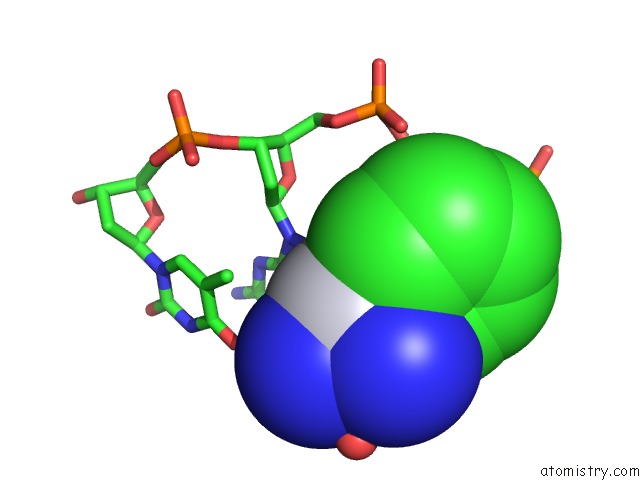
Mono view
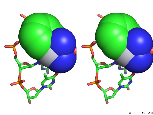
Stereo pair view
Mono view
Stereo pair view
A full contact list of Platinum with other atoms in the Pt binding
site number 1 of X-Ray Crystal Structure of A Monofunctional Platinum-Dna Adduct, Cis- {Pt(NH3)2(Pyridine)}2+ Bound to Deoxyguanosine in A Dodecamer Duplex within 5.0Å range:
|
Reference:
K.S.Lovejoy,
R.C.Todd,
S.Zhang,
M.S.Mccormick,
J.A.D'aquino,
J.T.Reardon,
A.Sancar,
K.M.Giacomini,
S.J.Lippard.
Cis-Diammine(Pyridine)Chloroplatinum(II), A Monofunctional Platinum(II) Antitumor Agent: Uptake, Structure, Function, and Prospects. Proc.Natl.Acad.Sci.Usa V. 105 8902 2008.
ISSN: ISSN 0027-8424
PubMed: 18579768
DOI: 10.1073/PNAS.0803441105
Page generated: Thu Oct 10 10:45:33 2024
ISSN: ISSN 0027-8424
PubMed: 18579768
DOI: 10.1073/PNAS.0803441105
Last articles
Zn in 9J0NZn in 9J0O
Zn in 9J0P
Zn in 9FJX
Zn in 9EKB
Zn in 9C0F
Zn in 9CAH
Zn in 9CH0
Zn in 9CH3
Zn in 9CH1