Platinum »
PDB 3ef3-3wua »
3tur »
Platinum in PDB 3tur: Crystal Structure of M. Tuberculosis Ld-Transpeptidase Type 2 Complexed with A Peptidoglycan Fragment
Protein crystallography data
The structure of Crystal Structure of M. Tuberculosis Ld-Transpeptidase Type 2 Complexed with A Peptidoglycan Fragment, PDB code: 3tur
was solved by
M.A.Bianchet,
S.B.Erdemli,
R.Gupta,
G.Lamichhane,
L.M.Amzel,
with X-Ray Crystallography technique. A brief refinement statistics is given in the table below:
| Resolution Low / High (Å) | 85.69 / 1.72 |
| Space group | I 21 21 21 |
| Cell size a, b, c (Å), α, β, γ (°) | 119.132, 120.829, 122.847, 90.00, 90.00, 90.00 |
| R / Rfree (%) | 19.9 / 23.5 |
Other elements in 3tur:
The structure of Crystal Structure of M. Tuberculosis Ld-Transpeptidase Type 2 Complexed with A Peptidoglycan Fragment also contains other interesting chemical elements:
| Iodine | (I) | 11 atoms |
Platinum Binding Sites:
Pages:
>>> Page 1 <<< Page 2, Binding sites: 11 - 12;Binding sites:
The binding sites of Platinum atom in the Crystal Structure of M. Tuberculosis Ld-Transpeptidase Type 2 Complexed with A Peptidoglycan Fragment (pdb code 3tur). This binding sites where shown within 5.0 Angstroms radius around Platinum atom.In total 12 binding sites of Platinum where determined in the Crystal Structure of M. Tuberculosis Ld-Transpeptidase Type 2 Complexed with A Peptidoglycan Fragment, PDB code: 3tur:
Jump to Platinum binding site number: 1; 2; 3; 4; 5; 6; 7; 8; 9; 10;
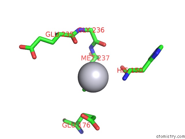
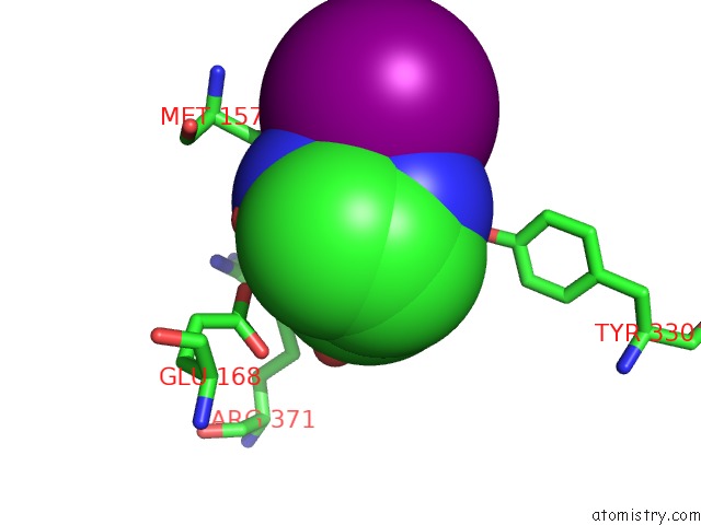
Platinum binding site 1 out of 12 in 3tur
Go back to
Platinum binding site 1 out
of 12 in the Crystal Structure of M. Tuberculosis Ld-Transpeptidase Type 2 Complexed with A Peptidoglycan Fragment
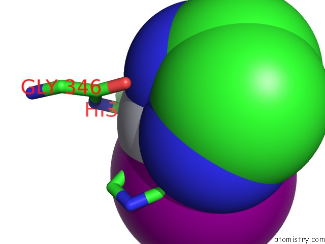
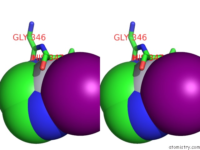
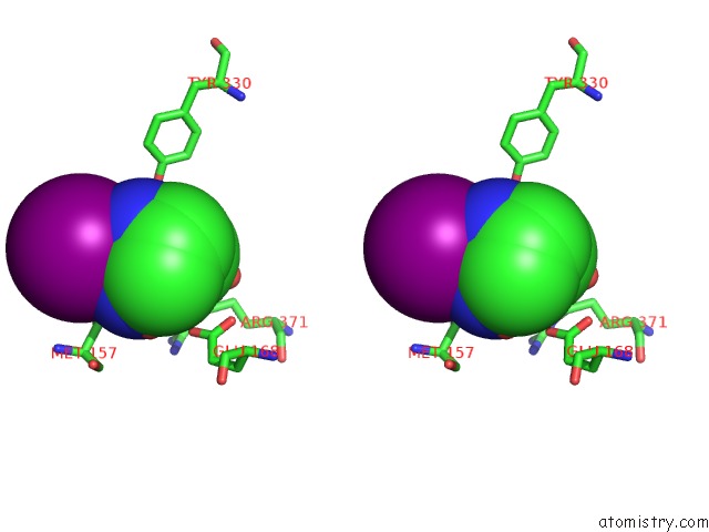
Mono view
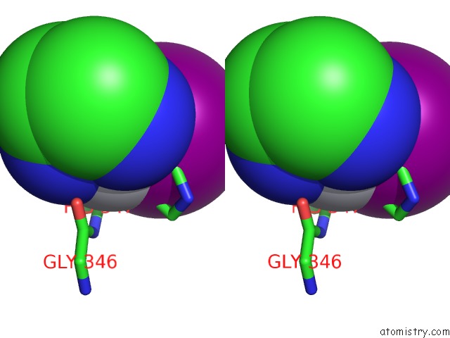
Stereo pair view
Mono view
Stereo pair view
A full contact list of Platinum with other atoms in the Pt binding
site number 1 of Crystal Structure of M. Tuberculosis Ld-Transpeptidase Type 2 Complexed with A Peptidoglycan Fragment within 5.0Å range:
|
Platinum binding site 2 out of 12 in 3tur
Go back to
Platinum binding site 2 out
of 12 in the Crystal Structure of M. Tuberculosis Ld-Transpeptidase Type 2 Complexed with A Peptidoglycan Fragment
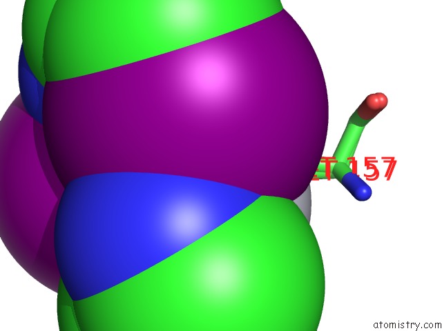
Mono view

Stereo pair view
Mono view
Stereo pair view
A full contact list of Platinum with other atoms in the Pt binding
site number 2 of Crystal Structure of M. Tuberculosis Ld-Transpeptidase Type 2 Complexed with A Peptidoglycan Fragment within 5.0Å range:
|
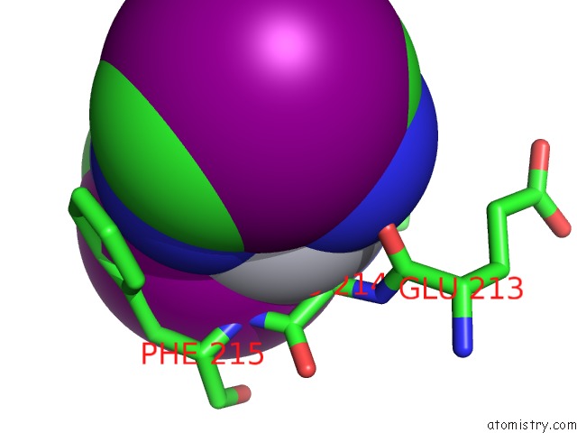
Platinum binding site 3 out of 12 in 3tur
Go back to
Platinum binding site 3 out
of 12 in the Crystal Structure of M. Tuberculosis Ld-Transpeptidase Type 2 Complexed with A Peptidoglycan Fragment

Mono view

Stereo pair view
Mono view
Stereo pair view
A full contact list of Platinum with other atoms in the Pt binding
site number 3 of Crystal Structure of M. Tuberculosis Ld-Transpeptidase Type 2 Complexed with A Peptidoglycan Fragment within 5.0Å range:
|
Platinum binding site 4 out of 12 in 3tur
Go back to
Platinum binding site 4 out
of 12 in the Crystal Structure of M. Tuberculosis Ld-Transpeptidase Type 2 Complexed with A Peptidoglycan Fragment

Mono view

Stereo pair view
Mono view
Stereo pair view
A full contact list of Platinum with other atoms in the Pt binding
site number 4 of Crystal Structure of M. Tuberculosis Ld-Transpeptidase Type 2 Complexed with A Peptidoglycan Fragment within 5.0Å range:
|
Platinum binding site 5 out of 12 in 3tur
Go back to
Platinum binding site 5 out
of 12 in the Crystal Structure of M. Tuberculosis Ld-Transpeptidase Type 2 Complexed with A Peptidoglycan Fragment

Mono view

Stereo pair view
Mono view
Stereo pair view
A full contact list of Platinum with other atoms in the Pt binding
site number 5 of Crystal Structure of M. Tuberculosis Ld-Transpeptidase Type 2 Complexed with A Peptidoglycan Fragment within 5.0Å range:
|
Platinum binding site 6 out of 12 in 3tur
Go back to
Platinum binding site 6 out
of 12 in the Crystal Structure of M. Tuberculosis Ld-Transpeptidase Type 2 Complexed with A Peptidoglycan Fragment
Mono view

Stereo pair view
Mono view
Stereo pair view
A full contact list of Platinum with other atoms in the Pt binding
site number 6 of Crystal Structure of M. Tuberculosis Ld-Transpeptidase Type 2 Complexed with A Peptidoglycan Fragment within 5.0Å range:
|
Platinum binding site 7 out of 12 in 3tur
Go back to
Platinum binding site 7 out
of 12 in the Crystal Structure of M. Tuberculosis Ld-Transpeptidase Type 2 Complexed with A Peptidoglycan Fragment
Mono view

Stereo pair view
Mono view
Stereo pair view
A full contact list of Platinum with other atoms in the Pt binding
site number 7 of Crystal Structure of M. Tuberculosis Ld-Transpeptidase Type 2 Complexed with A Peptidoglycan Fragment within 5.0Å range:
|
Platinum binding site 8 out of 12 in 3tur
Go back to
Platinum binding site 8 out
of 12 in the Crystal Structure of M. Tuberculosis Ld-Transpeptidase Type 2 Complexed with A Peptidoglycan Fragment

Mono view
Stereo pair view
Mono view
Stereo pair view
A full contact list of Platinum with other atoms in the Pt binding
site number 8 of Crystal Structure of M. Tuberculosis Ld-Transpeptidase Type 2 Complexed with A Peptidoglycan Fragment within 5.0Å range:
|
Platinum binding site 9 out of 12 in 3tur
Go back to
Platinum binding site 9 out
of 12 in the Crystal Structure of M. Tuberculosis Ld-Transpeptidase Type 2 Complexed with A Peptidoglycan Fragment

Mono view

Stereo pair view
Mono view
Stereo pair view
A full contact list of Platinum with other atoms in the Pt binding
site number 9 of Crystal Structure of M. Tuberculosis Ld-Transpeptidase Type 2 Complexed with A Peptidoglycan Fragment within 5.0Å range:
|
Platinum binding site 10 out of 12 in 3tur
Go back to
Platinum binding site 10 out
of 12 in the Crystal Structure of M. Tuberculosis Ld-Transpeptidase Type 2 Complexed with A Peptidoglycan Fragment
Mono view
Stereo pair view
Mono view
Stereo pair view
A full contact list of Platinum with other atoms in the Pt binding
site number 10 of Crystal Structure of M. Tuberculosis Ld-Transpeptidase Type 2 Complexed with A Peptidoglycan Fragment within 5.0Å range:
|
Reference:
S.B.Erdemli,
R.Gupta,
W.R.Bishai,
G.Lamichhane,
L.M.Amzel,
M.A.Bianchet.
Targeting the Cell Wall of Mycobacterium Tuberculosis: Structure and Mechanism of L,D-Transpeptidase 2. Structure V. 20 2103 2012.
ISSN: ISSN 0969-2126
PubMed: 23103390
DOI: 10.1016/J.STR.2012.09.016
Page generated: Thu Oct 10 10:49:33 2024
ISSN: ISSN 0969-2126
PubMed: 23103390
DOI: 10.1016/J.STR.2012.09.016
Last articles
Fe in 2YXOFe in 2YRS
Fe in 2YXC
Fe in 2YNM
Fe in 2YVJ
Fe in 2YP1
Fe in 2YU2
Fe in 2YU1
Fe in 2YQB
Fe in 2YOO