Platinum »
PDB 3ef3-3wua »
3txf »
Platinum in PDB 3txf: Hewl Co-Crystallization with Cisplatin in Dmso Media with Glycerol As the Cryoprotectant
Enzymatic activity of Hewl Co-Crystallization with Cisplatin in Dmso Media with Glycerol As the Cryoprotectant
All present enzymatic activity of Hewl Co-Crystallization with Cisplatin in Dmso Media with Glycerol As the Cryoprotectant:
3.2.1.17;
3.2.1.17;
Protein crystallography data
The structure of Hewl Co-Crystallization with Cisplatin in Dmso Media with Glycerol As the Cryoprotectant, PDB code: 3txf
was solved by
S.W.M.Tanley,
A.M.M.Schreurs,
J.R.Helliwell,
L.M.J.Kroon-Batenburg,
with X-Ray Crystallography technique. A brief refinement statistics is given in the table below:
| Resolution Low / High (Å) | 55.47 / 1.69 |
| Space group | P 43 21 2 |
| Cell size a, b, c (Å), α, β, γ (°) | 78.443, 78.443, 36.971, 90.00, 90.00, 90.00 |
| R / Rfree (%) | 17.5 / 24.9 |
Other elements in 3txf:
The structure of Hewl Co-Crystallization with Cisplatin in Dmso Media with Glycerol As the Cryoprotectant also contains other interesting chemical elements:
| Chlorine | (Cl) | 7 atoms |
| Sodium | (Na) | 1 atom |
Platinum Binding Sites:
The binding sites of Platinum atom in the Hewl Co-Crystallization with Cisplatin in Dmso Media with Glycerol As the Cryoprotectant
(pdb code 3txf). This binding sites where shown within
5.0 Angstroms radius around Platinum atom.
In total only one binding site of Platinum was determined in the Hewl Co-Crystallization with Cisplatin in Dmso Media with Glycerol As the Cryoprotectant, PDB code: 3txf:
In total only one binding site of Platinum was determined in the Hewl Co-Crystallization with Cisplatin in Dmso Media with Glycerol As the Cryoprotectant, PDB code: 3txf:
Platinum binding site 1 out of 1 in 3txf
Go back to
Platinum binding site 1 out
of 1 in the Hewl Co-Crystallization with Cisplatin in Dmso Media with Glycerol As the Cryoprotectant
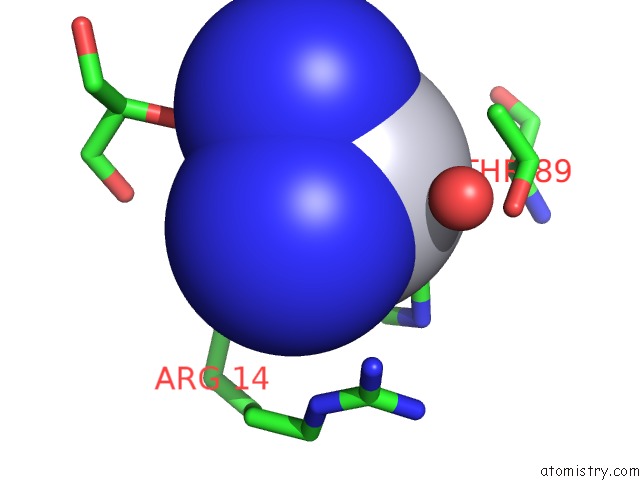
Mono view
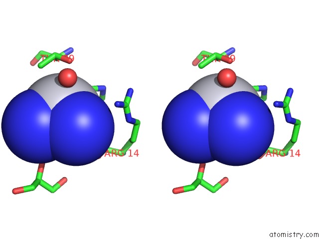
Stereo pair view
Mono view
Stereo pair view
A full contact list of Platinum with other atoms in the Pt binding
site number 1 of Hewl Co-Crystallization with Cisplatin in Dmso Media with Glycerol As the Cryoprotectant within 5.0Å range:
|
Reference:
S.W.Tanley,
A.M.Schreurs,
J.R.Helliwell,
L.M.Kroon-Batenburg.
Experience with Exchange and Archiving of Raw Data: Comparison of Data From Two Diffractometers and Four Software Packages on A Series of Lysozyme Crystals. J.Appl.Crystallogr. V. 46 108 2013.
ISSN: ISSN 0021-8898
PubMed: 23396873
DOI: 10.1107/S0021889812044172
Page generated: Thu Oct 10 10:50:03 2024
ISSN: ISSN 0021-8898
PubMed: 23396873
DOI: 10.1107/S0021889812044172
Last articles
Ca in 5LXSCa in 5LWD
Ca in 5LVD
Ca in 5LQ2
Ca in 5LW3
Ca in 5LPU
Ca in 5LUH
Ca in 5LUL
Ca in 5LU3
Ca in 5LSF