Platinum »
PDB 4omb-5djl »
4ot4 »
Platinum in PDB 4ot4: X-Ray Structure of the Adduct Formed Between Cisplatin and Ribonuclease A
Enzymatic activity of X-Ray Structure of the Adduct Formed Between Cisplatin and Ribonuclease A
All present enzymatic activity of X-Ray Structure of the Adduct Formed Between Cisplatin and Ribonuclease A:
3.1.27.5;
3.1.27.5;
Protein crystallography data
The structure of X-Ray Structure of the Adduct Formed Between Cisplatin and Ribonuclease A, PDB code: 4ot4
was solved by
L.Messori,
A.Merlino,
with X-Ray Crystallography technique. A brief refinement statistics is given in the table below:
| Resolution Low / High (Å) | 30.88 / 1.85 |
| Space group | C 1 2 1 |
| Cell size a, b, c (Å), α, β, γ (°) | 99.767, 32.463, 67.575, 90.00, 93.07, 90.00 |
| R / Rfree (%) | 18.9 / 24.7 |
Platinum Binding Sites:
The binding sites of Platinum atom in the X-Ray Structure of the Adduct Formed Between Cisplatin and Ribonuclease A
(pdb code 4ot4). This binding sites where shown within
5.0 Angstroms radius around Platinum atom.
In total 3 binding sites of Platinum where determined in the X-Ray Structure of the Adduct Formed Between Cisplatin and Ribonuclease A, PDB code: 4ot4:
Jump to Platinum binding site number: 1; 2; 3;
In total 3 binding sites of Platinum where determined in the X-Ray Structure of the Adduct Formed Between Cisplatin and Ribonuclease A, PDB code: 4ot4:
Jump to Platinum binding site number: 1; 2; 3;
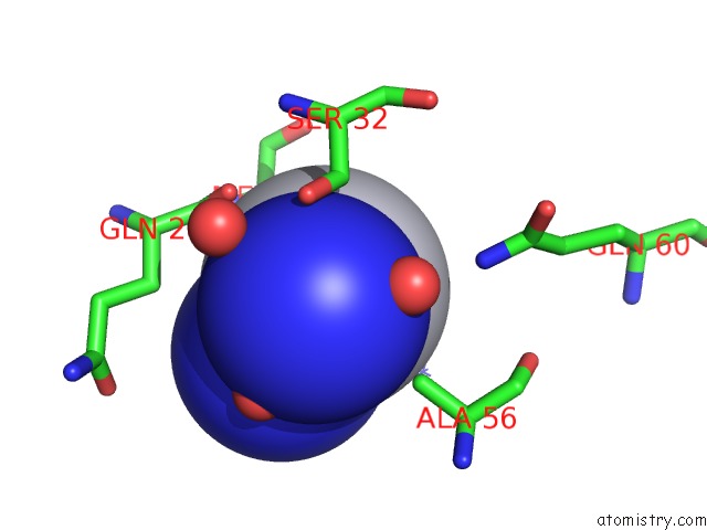
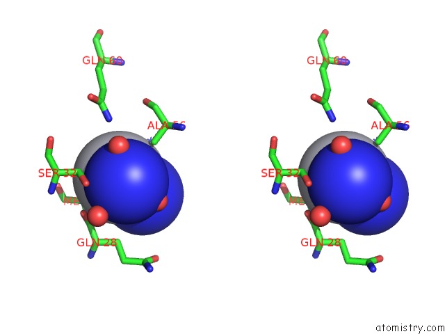
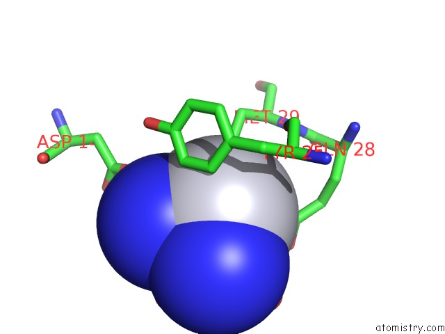
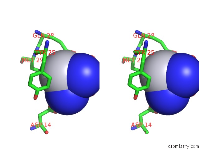
Platinum binding site 1 out of 3 in 4ot4
Go back to
Platinum binding site 1 out
of 3 in the X-Ray Structure of the Adduct Formed Between Cisplatin and Ribonuclease A
Mono view
Stereo pair view
Mono view
Stereo pair view
A full contact list of Platinum with other atoms in the Pt binding
site number 1 of X-Ray Structure of the Adduct Formed Between Cisplatin and Ribonuclease A within 5.0Å range:
|
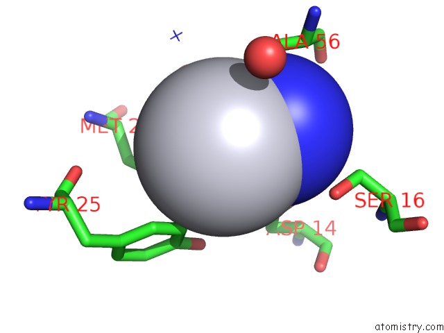
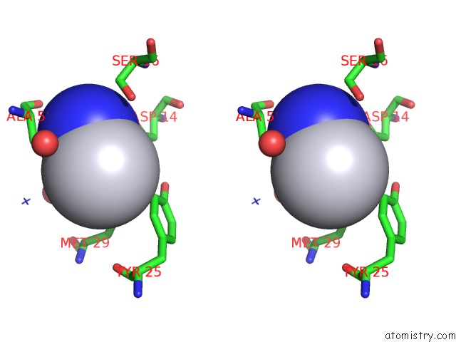
Platinum binding site 2 out of 3 in 4ot4
Go back to
Platinum binding site 2 out
of 3 in the X-Ray Structure of the Adduct Formed Between Cisplatin and Ribonuclease A
Mono view
Stereo pair view
Mono view
Stereo pair view
A full contact list of Platinum with other atoms in the Pt binding
site number 2 of X-Ray Structure of the Adduct Formed Between Cisplatin and Ribonuclease A within 5.0Å range:
|
Platinum binding site 3 out of 3 in 4ot4
Go back to
Platinum binding site 3 out
of 3 in the X-Ray Structure of the Adduct Formed Between Cisplatin and Ribonuclease A
Mono view
Stereo pair view
Mono view
Stereo pair view
A full contact list of Platinum with other atoms in the Pt binding
site number 3 of X-Ray Structure of the Adduct Formed Between Cisplatin and Ribonuclease A within 5.0Å range:
|
Reference:
L.Messori,
A.Merlino.
Cisplatin Binding to Proteins: Molecular Structure of the Ribonuclease A Adduct. Inorg.Chem. V. 53 3929 2014.
ISSN: ISSN 0020-1669
PubMed: 24694179
DOI: 10.1021/IC500360F
Page generated: Thu Oct 10 11:07:41 2024
ISSN: ISSN 0020-1669
PubMed: 24694179
DOI: 10.1021/IC500360F
Last articles
Fe in 2YXOFe in 2YRS
Fe in 2YXC
Fe in 2YNM
Fe in 2YVJ
Fe in 2YP1
Fe in 2YU2
Fe in 2YU1
Fe in 2YQB
Fe in 2YOO