Platinum »
PDB 4omb-5djl »
4pgw »
Platinum in PDB 4pgw: Crystal Structure of Yetj From Bacillus Subtilis at pH 6 By Pt-Sad
Protein crystallography data
The structure of Crystal Structure of Yetj From Bacillus Subtilis at pH 6 By Pt-Sad, PDB code: 4pgw
was solved by
Q.Liu,
Y.Chang,
W.A.Hendrickson,
New York Consortium On Membrane Proteinstructure (Nycomps),
with X-Ray Crystallography technique. A brief refinement statistics is given in the table below:
| Resolution Low / High (Å) | 36.77 / 3.60 |
| Space group | C 2 2 21 |
| Cell size a, b, c (Å), α, β, γ (°) | 47.640, 161.750, 123.830, 90.00, 90.00, 90.00 |
| R / Rfree (%) | 28.4 / 33 |
Platinum Binding Sites:
The binding sites of Platinum atom in the Crystal Structure of Yetj From Bacillus Subtilis at pH 6 By Pt-Sad
(pdb code 4pgw). This binding sites where shown within
5.0 Angstroms radius around Platinum atom.
In total 3 binding sites of Platinum where determined in the Crystal Structure of Yetj From Bacillus Subtilis at pH 6 By Pt-Sad, PDB code: 4pgw:
Jump to Platinum binding site number: 1; 2; 3;
In total 3 binding sites of Platinum where determined in the Crystal Structure of Yetj From Bacillus Subtilis at pH 6 By Pt-Sad, PDB code: 4pgw:
Jump to Platinum binding site number: 1; 2; 3;
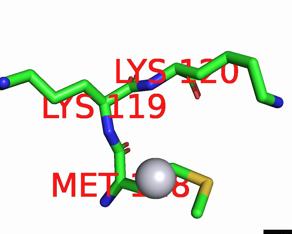
Platinum binding site 1 out of 3 in 4pgw
Go back to
Platinum binding site 1 out
of 3 in the Crystal Structure of Yetj From Bacillus Subtilis at pH 6 By Pt-Sad

Mono view

Stereo pair view
Mono view
Stereo pair view
A full contact list of Platinum with other atoms in the Pt binding
site number 1 of Crystal Structure of Yetj From Bacillus Subtilis at pH 6 By Pt-Sad within 5.0Å range:
|
Platinum binding site 2 out of 3 in 4pgw
Go back to
Platinum binding site 2 out
of 3 in the Crystal Structure of Yetj From Bacillus Subtilis at pH 6 By Pt-Sad

Mono view

Stereo pair view
Mono view
Stereo pair view
A full contact list of Platinum with other atoms in the Pt binding
site number 2 of Crystal Structure of Yetj From Bacillus Subtilis at pH 6 By Pt-Sad within 5.0Å range:
|
Platinum binding site 3 out of 3 in 4pgw
Go back to
Platinum binding site 3 out
of 3 in the Crystal Structure of Yetj From Bacillus Subtilis at pH 6 By Pt-Sad
Mono view

Stereo pair view
Mono view
Stereo pair view
A full contact list of Platinum with other atoms in the Pt binding
site number 3 of Crystal Structure of Yetj From Bacillus Subtilis at pH 6 By Pt-Sad within 5.0Å range:
|
Reference:
Y.Chang,
R.Bruni,
B.Kloss,
Z.Assur,
E.Kloppmann,
B.Rost,
W.A.Hendrickson,
Q.Liu.
Structural Basis For A pH-Sensitive Calcium Leak Across Membranes. Science V. 344 1131 2014.
ISSN: ESSN 1095-9203
PubMed: 24904158
DOI: 10.1126/SCIENCE.1252043
Page generated: Thu Oct 10 11:07:41 2024
ISSN: ESSN 1095-9203
PubMed: 24904158
DOI: 10.1126/SCIENCE.1252043
Last articles
Fe in 2YXOFe in 2YRS
Fe in 2YXC
Fe in 2YNM
Fe in 2YVJ
Fe in 2YP1
Fe in 2YU2
Fe in 2YU1
Fe in 2YQB
Fe in 2YOO