Platinum »
PDB 4omb-5djl »
4rsz »
Platinum in PDB 4rsz: The X-Ray Structure of the Primary Adduct Formed in the Reaction Between Cisplatin and Cytochrome C
Protein crystallography data
The structure of The X-Ray Structure of the Primary Adduct Formed in the Reaction Between Cisplatin and Cytochrome C, PDB code: 4rsz
was solved by
A.Merlino,
with X-Ray Crystallography technique. A brief refinement statistics is given in the table below:
| Resolution Low / High (Å) | 104.22 / 2.19 |
| Space group | P 3 |
| Cell size a, b, c (Å), α, β, γ (°) | 120.344, 120.344, 36.673, 90.00, 90.00, 120.00 |
| R / Rfree (%) | 23.1 / 28.2 |
Other elements in 4rsz:
The structure of The X-Ray Structure of the Primary Adduct Formed in the Reaction Between Cisplatin and Cytochrome C also contains other interesting chemical elements:
| Iron | (Fe) | 6 atoms |
| Chlorine | (Cl) | 3 atoms |
Platinum Binding Sites:
The binding sites of Platinum atom in the The X-Ray Structure of the Primary Adduct Formed in the Reaction Between Cisplatin and Cytochrome C
(pdb code 4rsz). This binding sites where shown within
5.0 Angstroms radius around Platinum atom.
In total 5 binding sites of Platinum where determined in the The X-Ray Structure of the Primary Adduct Formed in the Reaction Between Cisplatin and Cytochrome C, PDB code: 4rsz:
Jump to Platinum binding site number: 1; 2; 3; 4; 5;
In total 5 binding sites of Platinum where determined in the The X-Ray Structure of the Primary Adduct Formed in the Reaction Between Cisplatin and Cytochrome C, PDB code: 4rsz:
Jump to Platinum binding site number: 1; 2; 3; 4; 5;
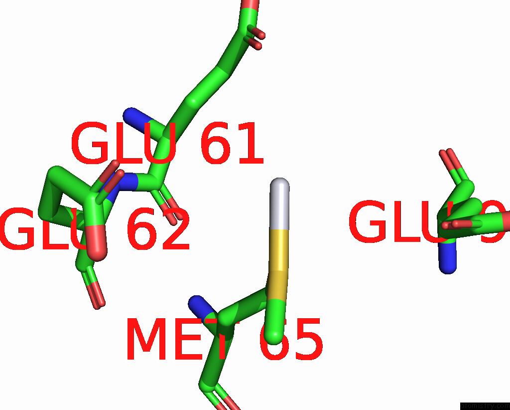
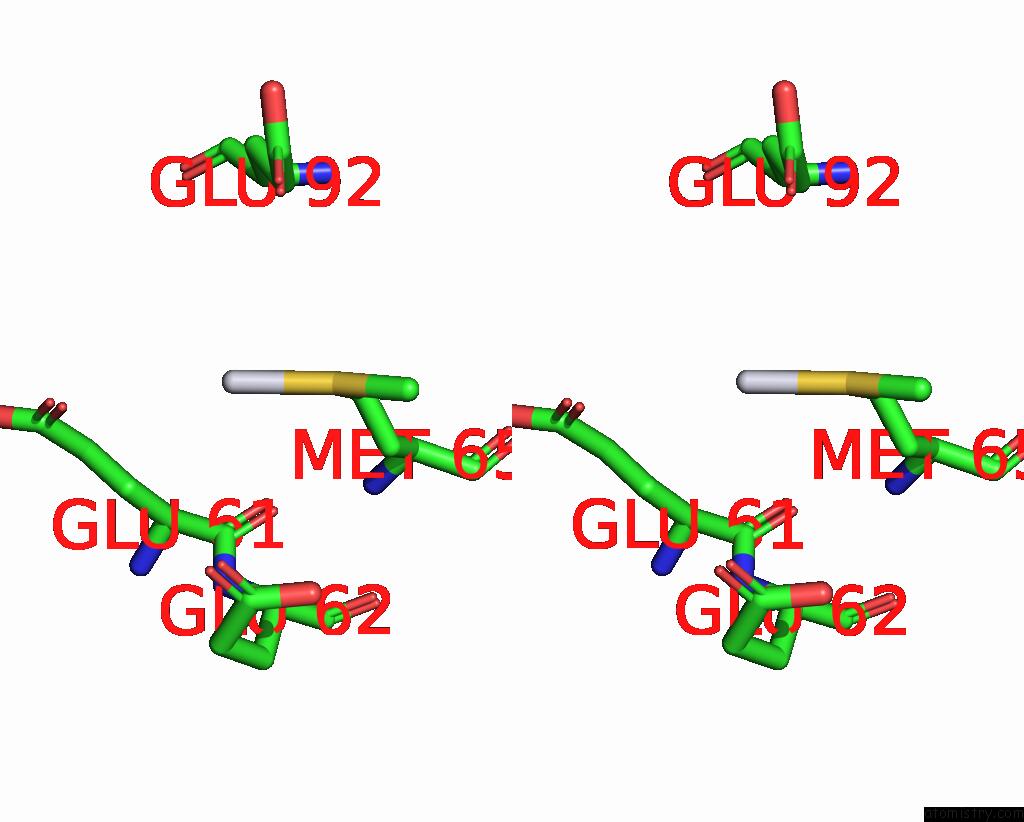
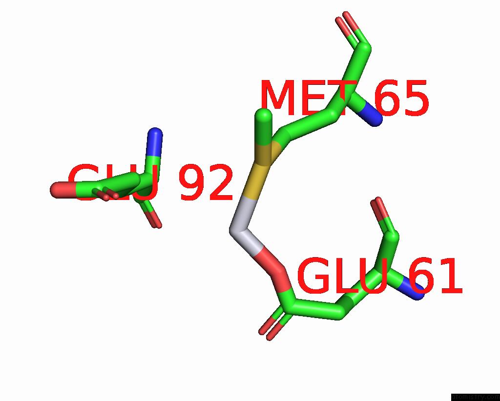
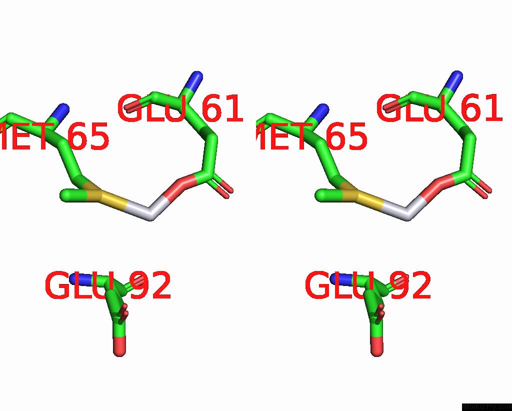
Platinum binding site 1 out of 5 in 4rsz
Go back to
Platinum binding site 1 out
of 5 in the The X-Ray Structure of the Primary Adduct Formed in the Reaction Between Cisplatin and Cytochrome C
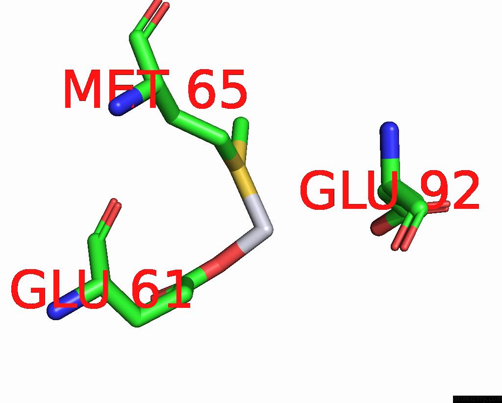
Mono view
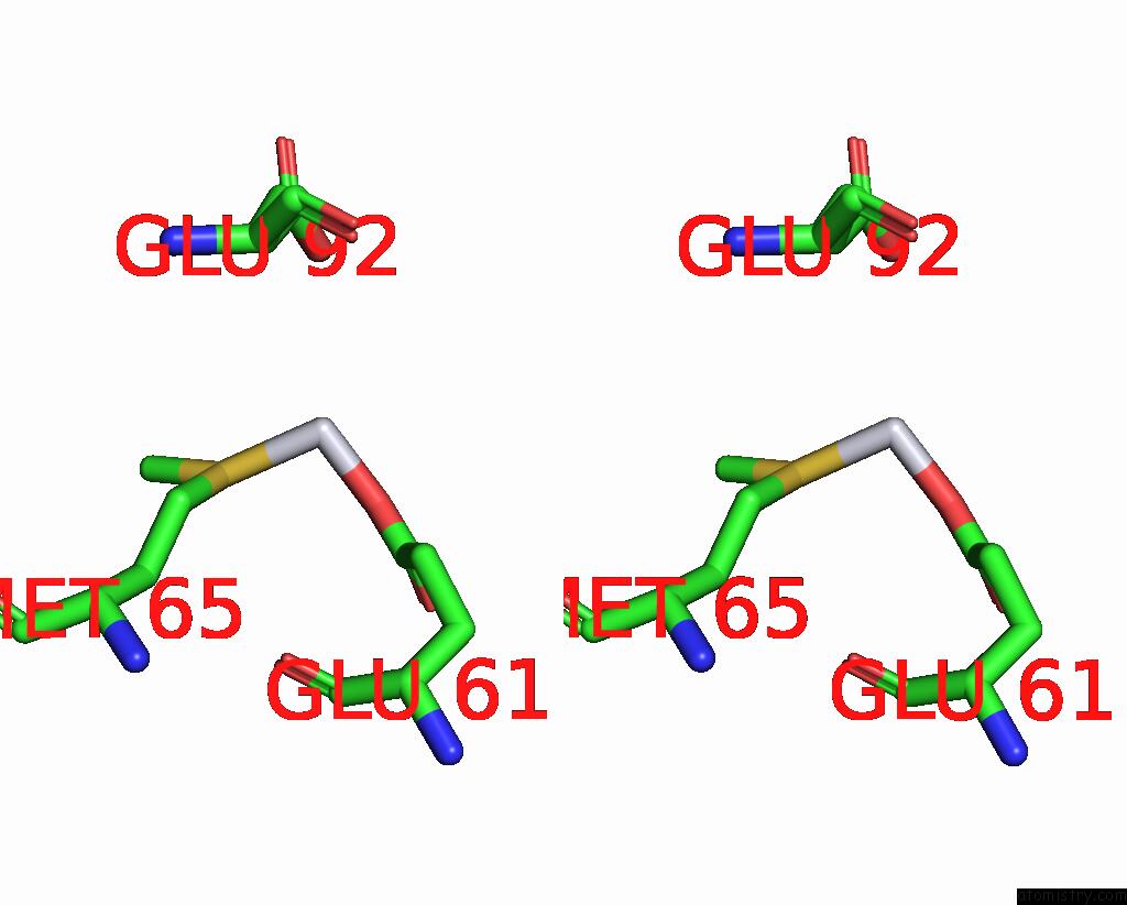
Stereo pair view
Mono view
Stereo pair view
A full contact list of Platinum with other atoms in the Pt binding
site number 1 of The X-Ray Structure of the Primary Adduct Formed in the Reaction Between Cisplatin and Cytochrome C within 5.0Å range:
|
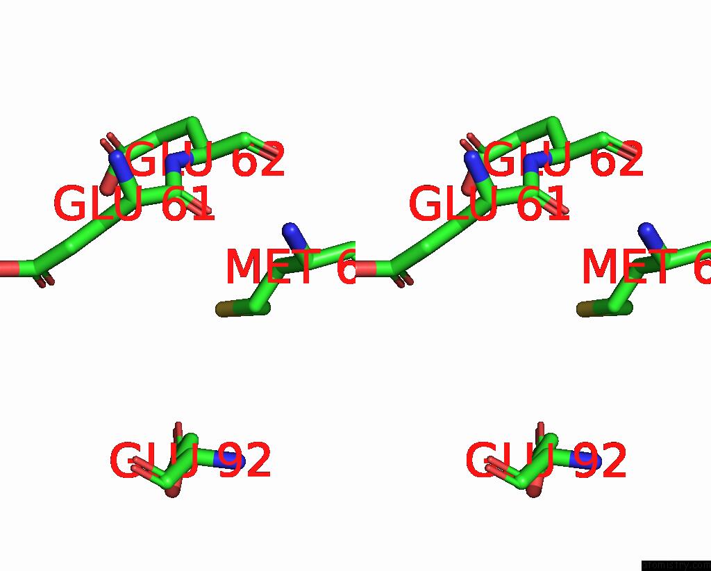
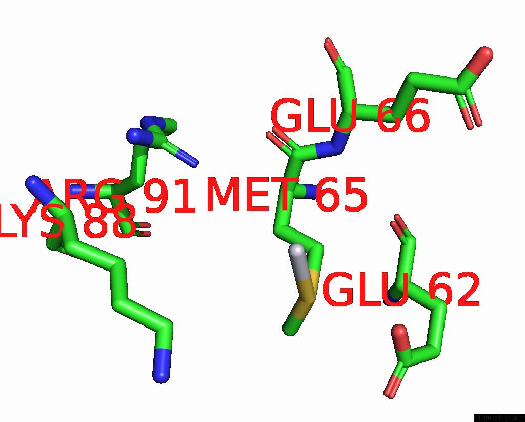
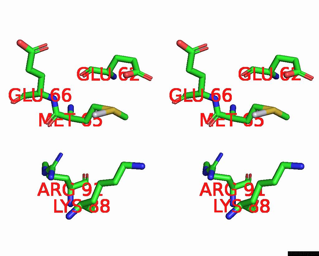
Platinum binding site 2 out of 5 in 4rsz
Go back to
Platinum binding site 2 out
of 5 in the The X-Ray Structure of the Primary Adduct Formed in the Reaction Between Cisplatin and Cytochrome C
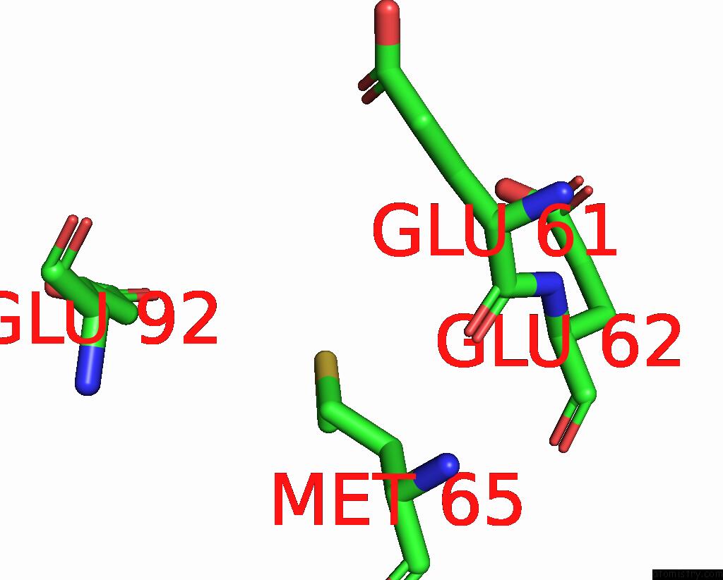
Mono view
Stereo pair view
Mono view
Stereo pair view
A full contact list of Platinum with other atoms in the Pt binding
site number 2 of The X-Ray Structure of the Primary Adduct Formed in the Reaction Between Cisplatin and Cytochrome C within 5.0Å range:
|
Platinum binding site 3 out of 5 in 4rsz
Go back to
Platinum binding site 3 out
of 5 in the The X-Ray Structure of the Primary Adduct Formed in the Reaction Between Cisplatin and Cytochrome C
Mono view
Stereo pair view
Mono view
Stereo pair view
A full contact list of Platinum with other atoms in the Pt binding
site number 3 of The X-Ray Structure of the Primary Adduct Formed in the Reaction Between Cisplatin and Cytochrome C within 5.0Å range:
|
Platinum binding site 4 out of 5 in 4rsz
Go back to
Platinum binding site 4 out
of 5 in the The X-Ray Structure of the Primary Adduct Formed in the Reaction Between Cisplatin and Cytochrome C
Mono view
Stereo pair view
Mono view
Stereo pair view
A full contact list of Platinum with other atoms in the Pt binding
site number 4 of The X-Ray Structure of the Primary Adduct Formed in the Reaction Between Cisplatin and Cytochrome C within 5.0Å range:
|
Platinum binding site 5 out of 5 in 4rsz
Go back to
Platinum binding site 5 out
of 5 in the The X-Ray Structure of the Primary Adduct Formed in the Reaction Between Cisplatin and Cytochrome C
Mono view
Stereo pair view
Mono view
Stereo pair view
A full contact list of Platinum with other atoms in the Pt binding
site number 5 of The X-Ray Structure of the Primary Adduct Formed in the Reaction Between Cisplatin and Cytochrome C within 5.0Å range:
|
Reference:
G.Ferraro,
L.Messori,
A.Merlino.
The X-Ray Structure of the Primary Adducts Formed in the Reaction Between Cisplatin and Cytochrome C Chem.Commun.(Camb.) 2014.
ISSN: ESSN 1364-548X
DOI: 10.1039/C4CC09056J
Page generated: Thu Oct 10 11:09:51 2024
ISSN: ESSN 1364-548X
DOI: 10.1039/C4CC09056J
Last articles
Ca in 5MHLCa in 5MHC
Ca in 5MFL
Ca in 5MFN
Ca in 5MFH
Ca in 5MFE
Ca in 5MFG
Ca in 5MFA
Ca in 5MF4
Ca in 5MEY