Platinum »
PDB 1a2e-1sj7 »
1qnq »
Platinum in PDB 1qnq: The 3-D Structure of A Trichoderma Reesei B-Mannanase From Glycoside Hydrolase Family 5
Enzymatic activity of The 3-D Structure of A Trichoderma Reesei B-Mannanase From Glycoside Hydrolase Family 5
All present enzymatic activity of The 3-D Structure of A Trichoderma Reesei B-Mannanase From Glycoside Hydrolase Family 5:
3.2.1.78;
3.2.1.78;
Protein crystallography data
The structure of The 3-D Structure of A Trichoderma Reesei B-Mannanase From Glycoside Hydrolase Family 5, PDB code: 1qnq
was solved by
E.Sabini,
H.Schubert,
G.Murshudov,
K.S.Wilson,
M.Siika-Aho,
M.Penttila,
with X-Ray Crystallography technique. A brief refinement statistics is given in the table below:
| Resolution Low / High (Å) | 20.00 / 1.65 |
| Space group | P 1 21 1 |
| Cell size a, b, c (Å), α, β, γ (°) | 51.130, 54.290, 61.050, 90.00, 110.21, 90.00 |
| R / Rfree (%) | 11.5 / 16 |
Platinum Binding Sites:
The binding sites of Platinum atom in the The 3-D Structure of A Trichoderma Reesei B-Mannanase From Glycoside Hydrolase Family 5
(pdb code 1qnq). This binding sites where shown within
5.0 Angstroms radius around Platinum atom.
In total 2 binding sites of Platinum where determined in the The 3-D Structure of A Trichoderma Reesei B-Mannanase From Glycoside Hydrolase Family 5, PDB code: 1qnq:
Jump to Platinum binding site number: 1; 2;
In total 2 binding sites of Platinum where determined in the The 3-D Structure of A Trichoderma Reesei B-Mannanase From Glycoside Hydrolase Family 5, PDB code: 1qnq:
Jump to Platinum binding site number: 1; 2;
Platinum binding site 1 out of 2 in 1qnq
Go back to Platinum Binding Sites List in 1qnq
Platinum binding site 1 out
of 2 in the The 3-D Structure of A Trichoderma Reesei B-Mannanase From Glycoside Hydrolase Family 5
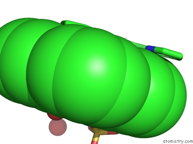
Mono view
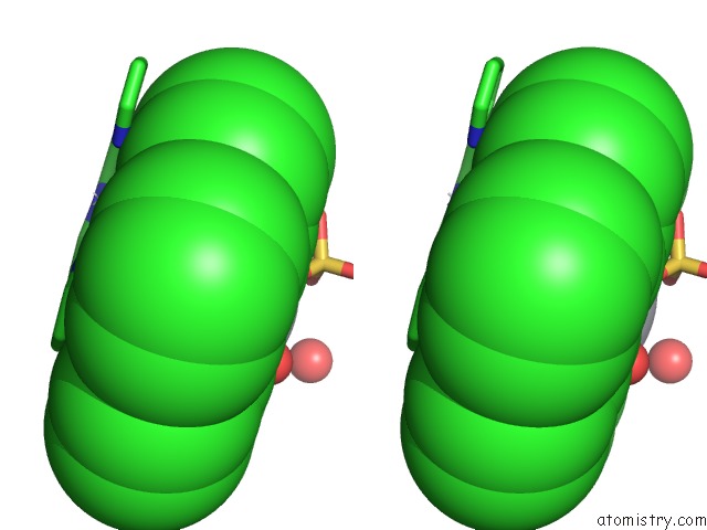
Stereo pair view
Mono view
Stereo pair view
A full contact list of Platinum with other atoms in the Pt binding
site number 1 of The 3-D Structure of A Trichoderma Reesei B-Mannanase From Glycoside Hydrolase Family 5 within 5.0Å range:
|
Platinum binding site 2 out of 2 in 1qnq
Go back to
Platinum Binding Sites List in 1qnq
Platinum binding site 2 out
of 2 in the The 3-D Structure of A Trichoderma Reesei B-Mannanase From Glycoside Hydrolase Family 5
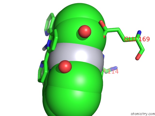
Mono view
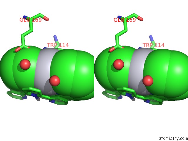
Stereo pair view
Mono view
Stereo pair view
A full contact list of Platinum with other atoms in the Pt binding
site number 2 of The 3-D Structure of A Trichoderma Reesei B-Mannanase From Glycoside Hydrolase Family 5 within 5.0Å range:
|
Reference:
E.Sabini,
H.Schubert,
G.Murshudov,
K.S.Wilson,
M.Siika-Aho,
M.Penttila.
The Three-Dimensional Structure of A Trichoderma Reesei Beta-Mannanase From Glycoside Hydrolase Family 5. Acta Crystallogr.,Sect.D V. 56 3 2000.
ISSN: ISSN 0907-4449
PubMed: 10666621
DOI: 10.1107/S0907444999013943
Page generated: Thu Oct 10 10:36:42 2024
ISSN: ISSN 0907-4449
PubMed: 10666621
DOI: 10.1107/S0907444999013943
Last articles
Cl in 3TY1Cl in 3TXK
Cl in 3TYT
Cl in 3TZ4
Cl in 3TZ0
Cl in 3TXG
Cl in 3TYK
Cl in 3TXI
Cl in 3TXJ
Cl in 3TXH
