Platinum »
PDB 1uas-3co3 »
2fyf »
Platinum in PDB 2fyf: Structure of A Putative Phosphoserine Aminotransferase From Mycobacterium Tuberculosis
Enzymatic activity of Structure of A Putative Phosphoserine Aminotransferase From Mycobacterium Tuberculosis
All present enzymatic activity of Structure of A Putative Phosphoserine Aminotransferase From Mycobacterium Tuberculosis:
2.6.1.52;
2.6.1.52;
Protein crystallography data
The structure of Structure of A Putative Phosphoserine Aminotransferase From Mycobacterium Tuberculosis, PDB code: 2fyf
was solved by
F.Coulibaly,
E.Lassalle,
E.N.Baker,
Mycobacterium Tuberculosisstructural Proteomics Project (Xmtb),
with X-Ray Crystallography technique. A brief refinement statistics is given in the table below:
| Resolution Low / High (Å) | 19.93 / 1.50 |
| Space group | P 21 21 21 |
| Cell size a, b, c (Å), α, β, γ (°) | 77.478, 94.056, 101.063, 90.00, 90.00, 90.00 |
| R / Rfree (%) | 14.4 / 16.6 |
Other elements in 2fyf:
The structure of Structure of A Putative Phosphoserine Aminotransferase From Mycobacterium Tuberculosis also contains other interesting chemical elements:
| Chlorine | (Cl) | 9 atoms |
Platinum Binding Sites:
The binding sites of Platinum atom in the Structure of A Putative Phosphoserine Aminotransferase From Mycobacterium Tuberculosis
(pdb code 2fyf). This binding sites where shown within
5.0 Angstroms radius around Platinum atom.
In total 6 binding sites of Platinum where determined in the Structure of A Putative Phosphoserine Aminotransferase From Mycobacterium Tuberculosis, PDB code: 2fyf:
Jump to Platinum binding site number: 1; 2; 3; 4; 5; 6;
In total 6 binding sites of Platinum where determined in the Structure of A Putative Phosphoserine Aminotransferase From Mycobacterium Tuberculosis, PDB code: 2fyf:
Jump to Platinum binding site number: 1; 2; 3; 4; 5; 6;
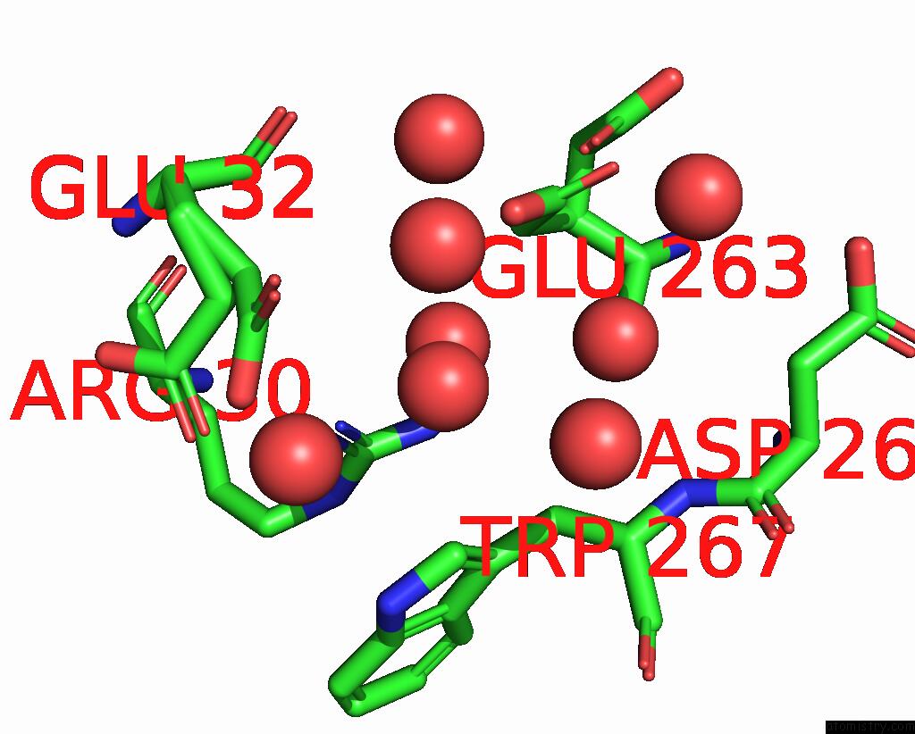
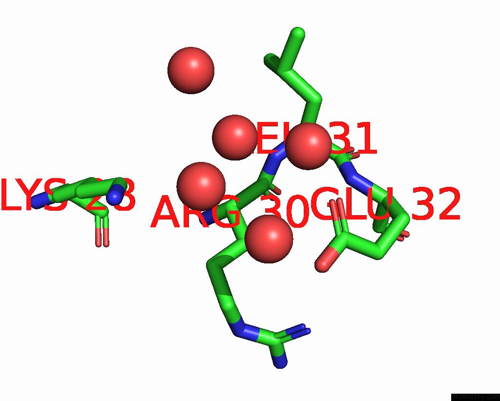
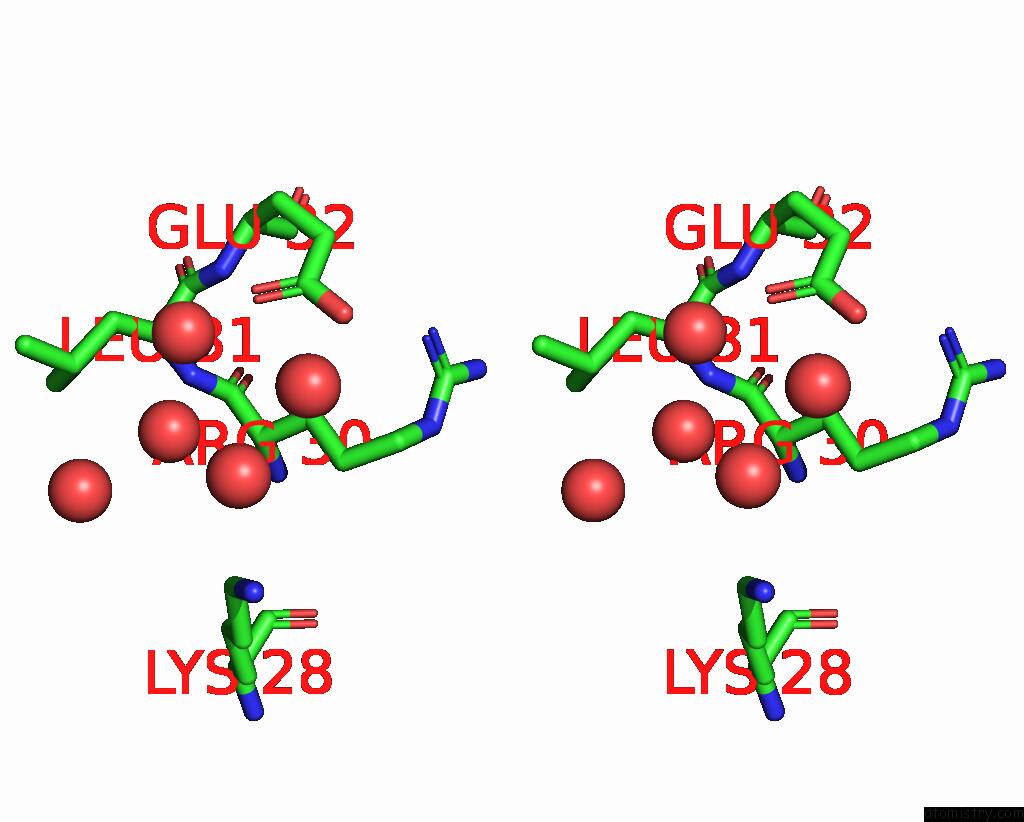
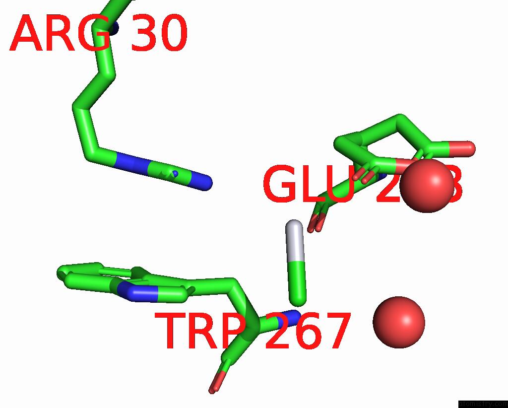
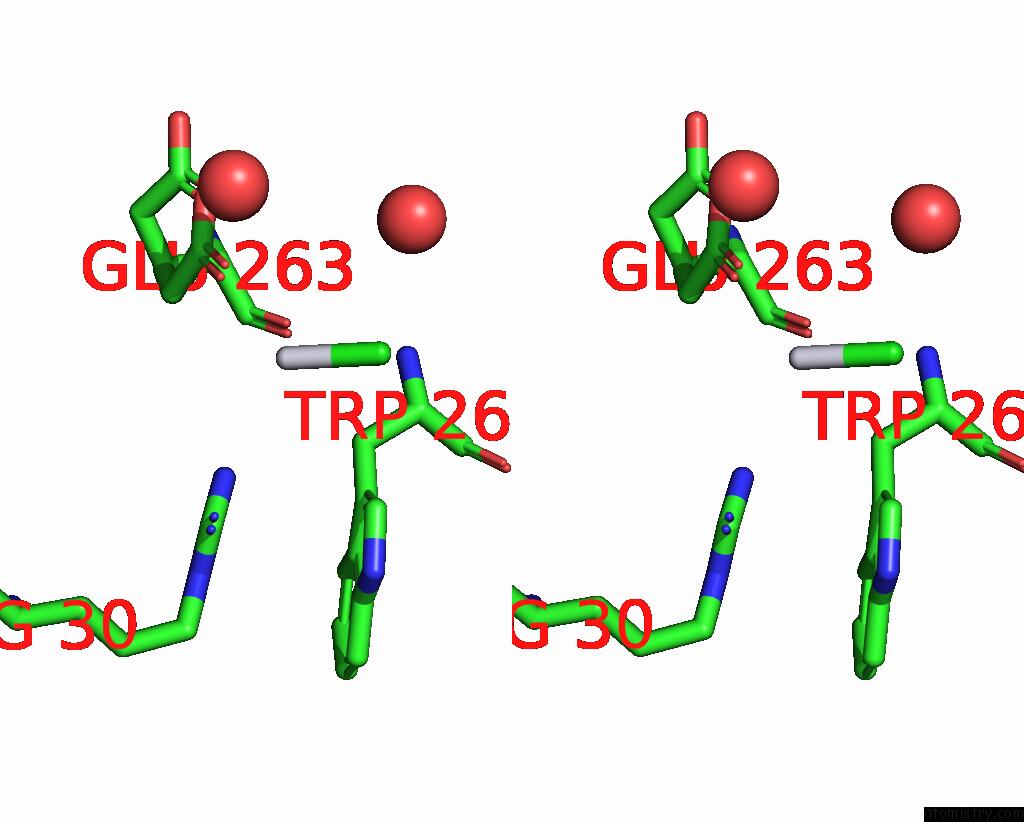
Platinum binding site 1 out of 6 in 2fyf
Go back to
Platinum binding site 1 out
of 6 in the Structure of A Putative Phosphoserine Aminotransferase From Mycobacterium Tuberculosis
Mono view
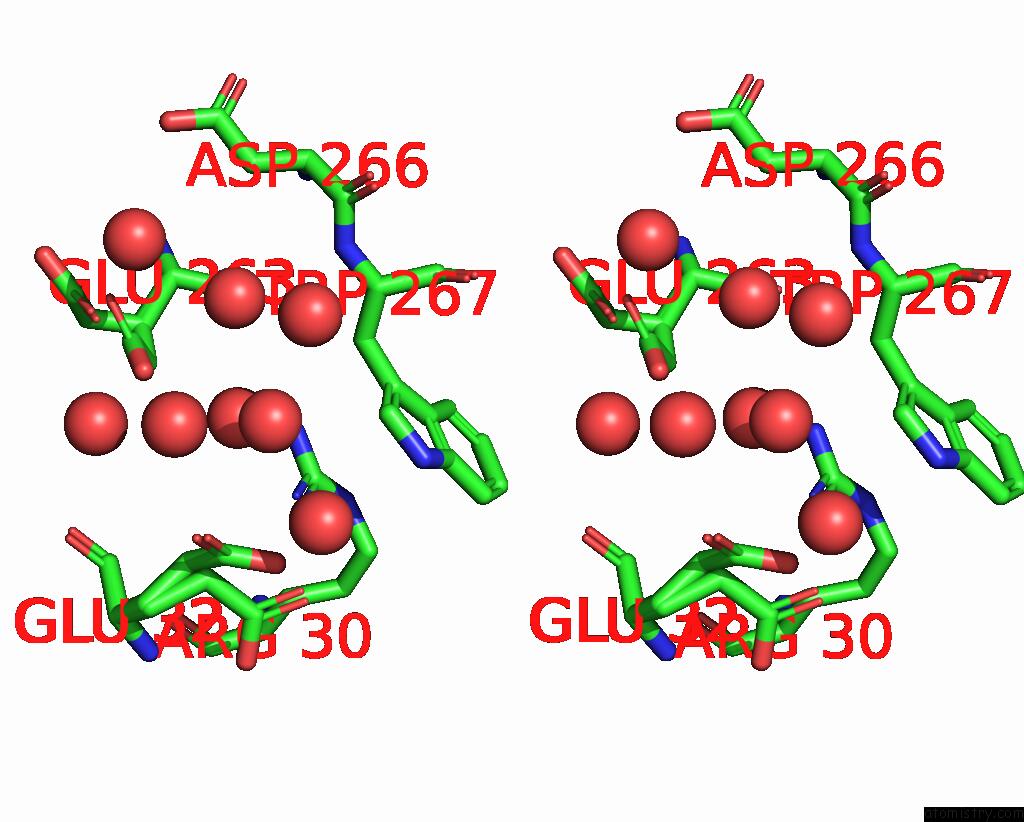
Stereo pair view
Mono view
Stereo pair view
A full contact list of Platinum with other atoms in the Pt binding
site number 1 of Structure of A Putative Phosphoserine Aminotransferase From Mycobacterium Tuberculosis within 5.0Å range:
|
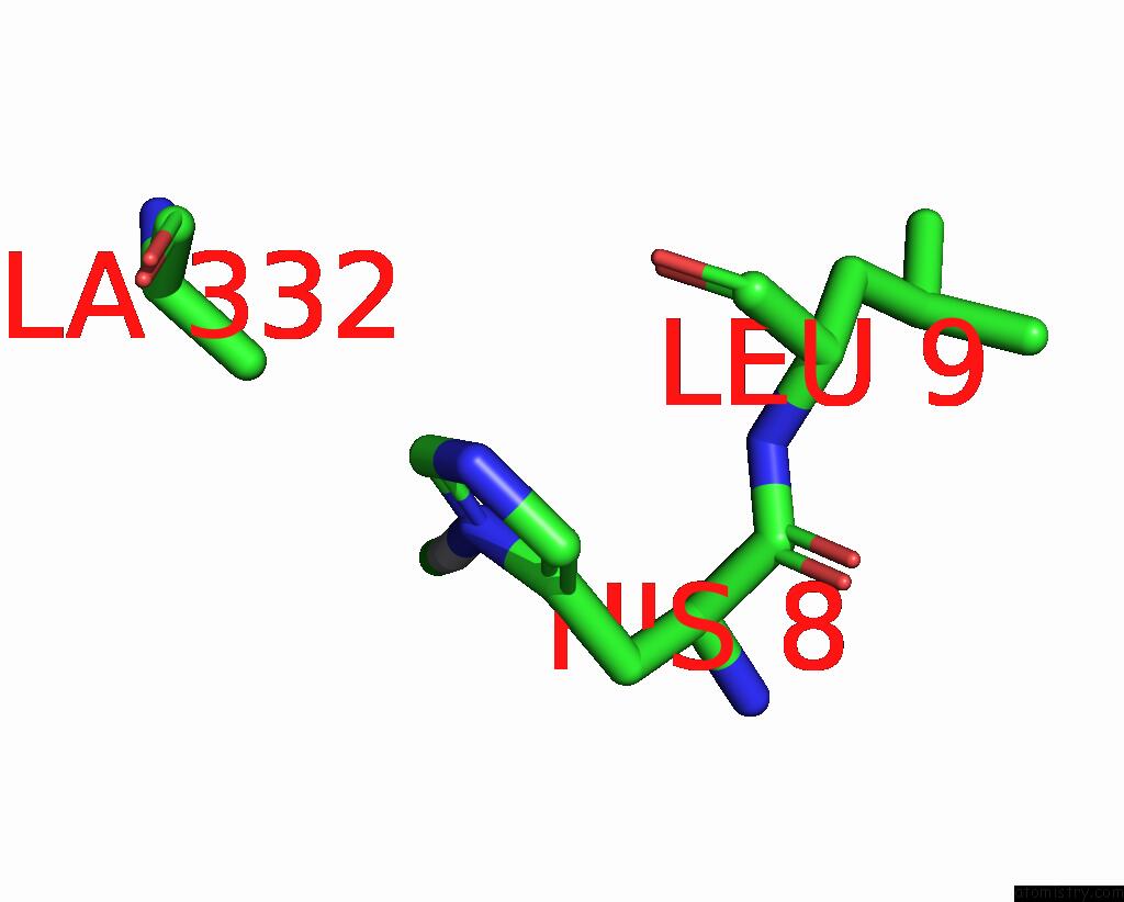
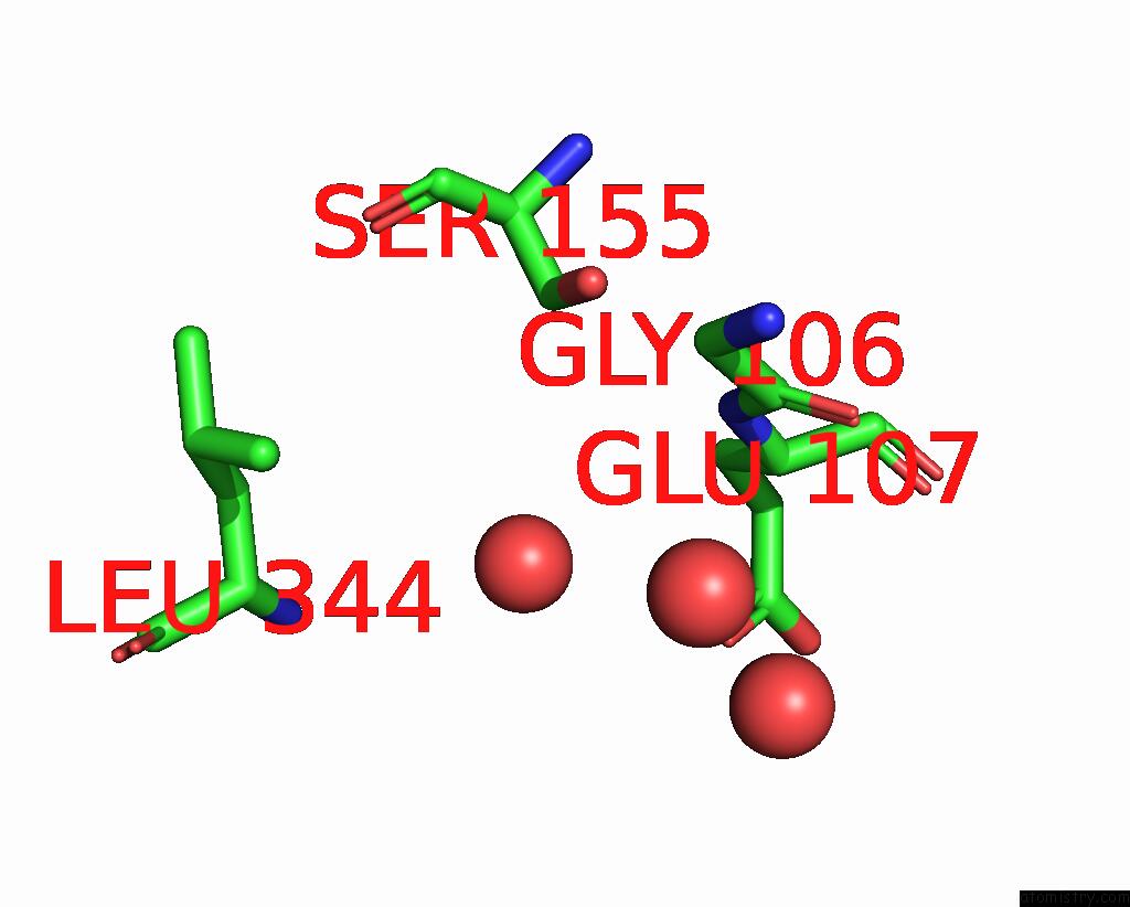
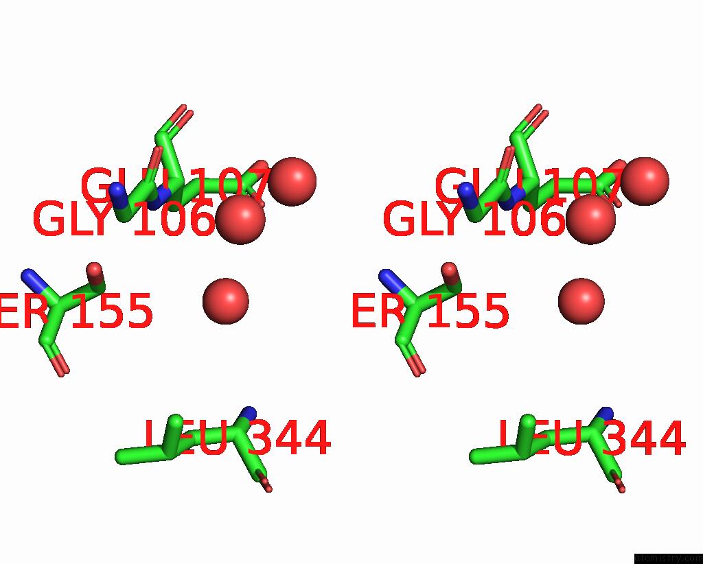
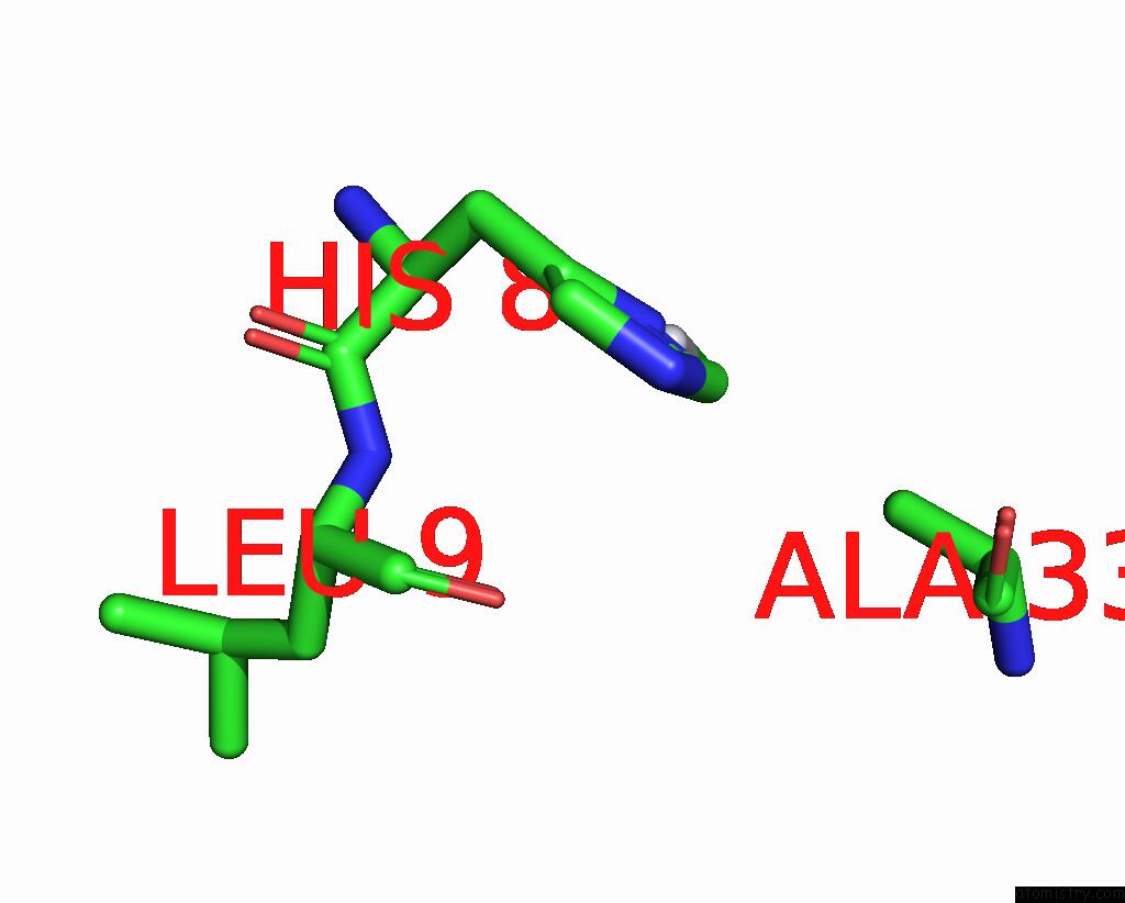
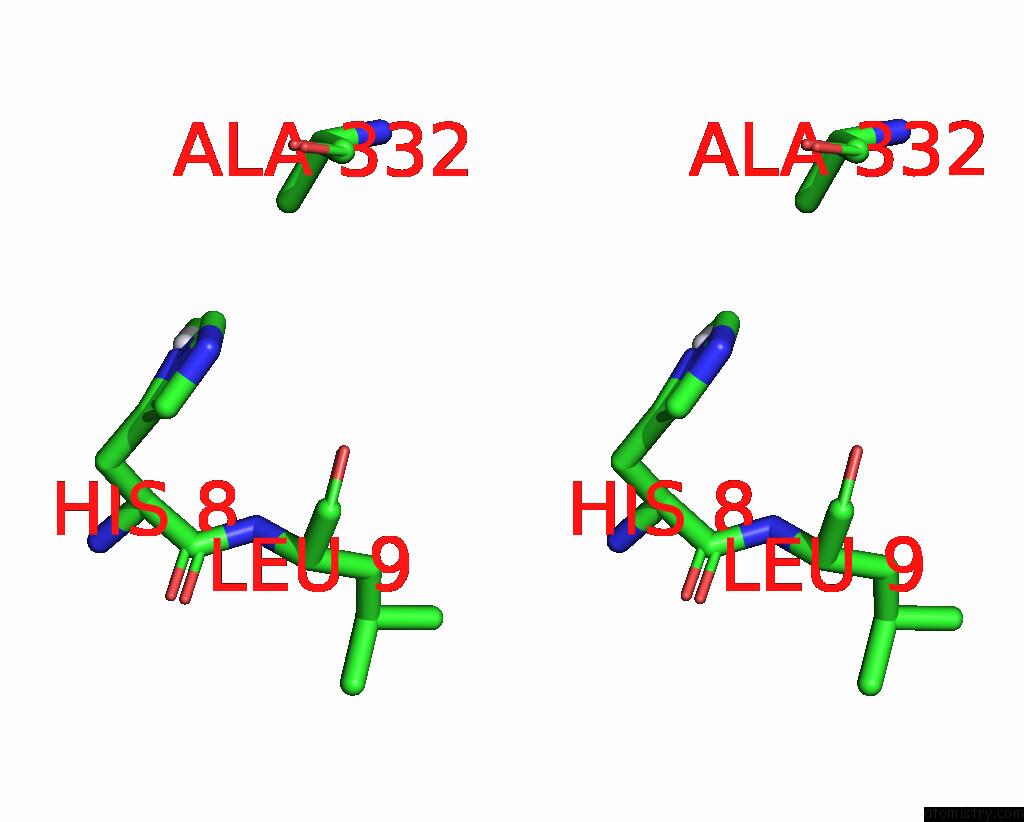
Platinum binding site 2 out of 6 in 2fyf
Go back to
Platinum binding site 2 out
of 6 in the Structure of A Putative Phosphoserine Aminotransferase From Mycobacterium Tuberculosis
Mono view
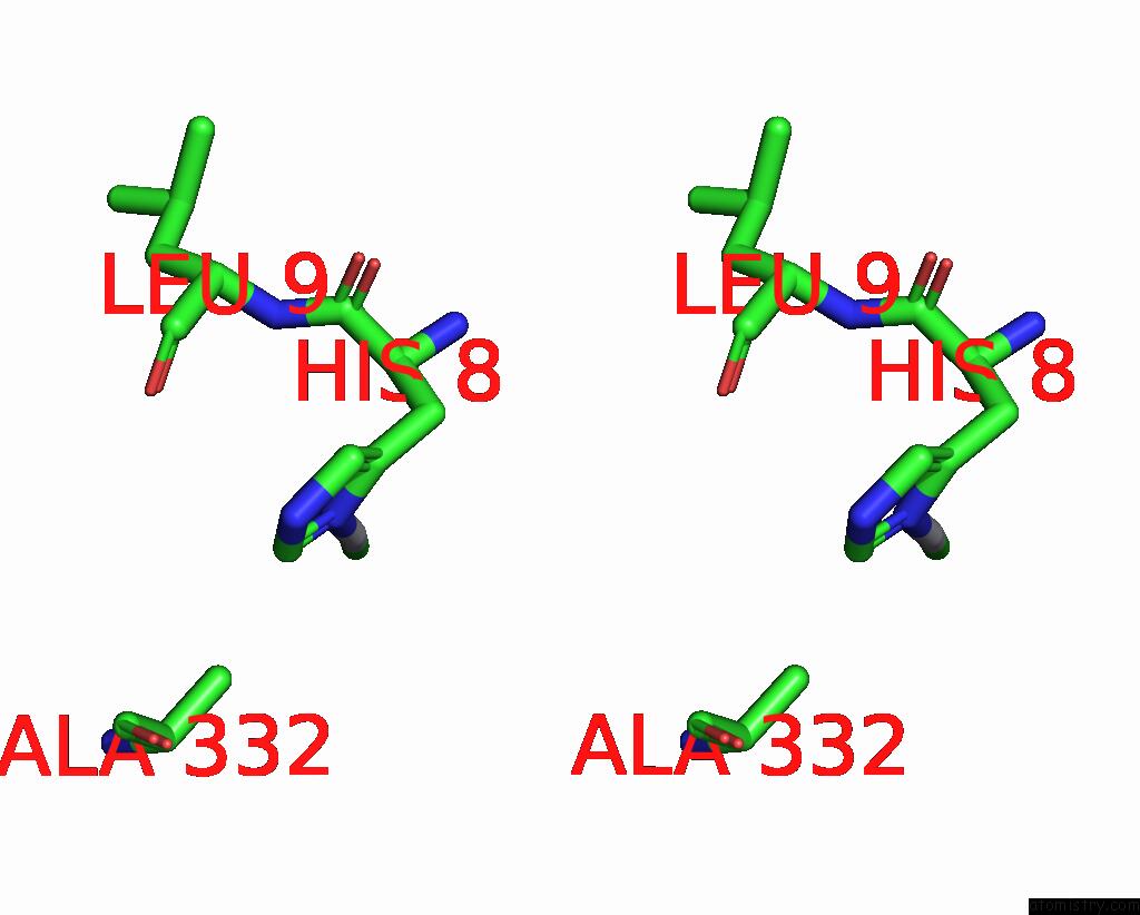
Stereo pair view
Mono view
Stereo pair view
A full contact list of Platinum with other atoms in the Pt binding
site number 2 of Structure of A Putative Phosphoserine Aminotransferase From Mycobacterium Tuberculosis within 5.0Å range:
|
Platinum binding site 3 out of 6 in 2fyf
Go back to
Platinum binding site 3 out
of 6 in the Structure of A Putative Phosphoserine Aminotransferase From Mycobacterium Tuberculosis
Mono view
Stereo pair view
Mono view
Stereo pair view
A full contact list of Platinum with other atoms in the Pt binding
site number 3 of Structure of A Putative Phosphoserine Aminotransferase From Mycobacterium Tuberculosis within 5.0Å range:
|
Platinum binding site 4 out of 6 in 2fyf
Go back to
Platinum binding site 4 out
of 6 in the Structure of A Putative Phosphoserine Aminotransferase From Mycobacterium Tuberculosis
Mono view
Stereo pair view
Mono view
Stereo pair view
A full contact list of Platinum with other atoms in the Pt binding
site number 4 of Structure of A Putative Phosphoserine Aminotransferase From Mycobacterium Tuberculosis within 5.0Å range:
|
Platinum binding site 5 out of 6 in 2fyf
Go back to
Platinum binding site 5 out
of 6 in the Structure of A Putative Phosphoserine Aminotransferase From Mycobacterium Tuberculosis
Mono view
Stereo pair view
Mono view
Stereo pair view
A full contact list of Platinum with other atoms in the Pt binding
site number 5 of Structure of A Putative Phosphoserine Aminotransferase From Mycobacterium Tuberculosis within 5.0Å range:
|
Platinum binding site 6 out of 6 in 2fyf
Go back to
Platinum binding site 6 out
of 6 in the Structure of A Putative Phosphoserine Aminotransferase From Mycobacterium Tuberculosis
Mono view
Stereo pair view
Mono view
Stereo pair view
A full contact list of Platinum with other atoms in the Pt binding
site number 6 of Structure of A Putative Phosphoserine Aminotransferase From Mycobacterium Tuberculosis within 5.0Å range:
|
Reference:
F.Coulibaly,
E.Lassalle,
H.M.Baker,
E.N.Baker.
Structure of Phosphoserine Aminotransferase From Mycobacterium Tuberculosis. Acta Crystallogr.,Sect.D V. 68 553 2012.
ISSN: ISSN 0907-4449
PubMed: 22525753
DOI: 10.1107/S0907444912004829
Page generated: Thu Oct 10 10:41:57 2024
ISSN: ISSN 0907-4449
PubMed: 22525753
DOI: 10.1107/S0907444912004829
Last articles
K in 4RN0K in 4RLG
K in 4RGQ
K in 4RKJ
K in 4RJ9
K in 4RJZ
K in 4RES
K in 4RDQ
K in 4RFL
K in 4R44