Platinum »
PDB 7dis-9h49 »
8auc »
Platinum in PDB 8auc: Structure of Peptidoglycan Hydrolase CG1735 From Corynebacterium Glutamicum, Trigonal Crystal Form
Protein crystallography data
The structure of Structure of Peptidoglycan Hydrolase CG1735 From Corynebacterium Glutamicum, Trigonal Crystal Form, PDB code: 8auc
was solved by
Q.Gaday,
A.M.Wehenkel,
P.M.Alzari,
P.Legrand,
with X-Ray Crystallography technique. A brief refinement statistics is given in the table below:
| Resolution Low / High (Å) | 49.57 / 3.50 |
| Space group | H 3 2 |
| Cell size a, b, c (Å), α, β, γ (°) | 230.58, 230.58, 416.69, 90, 90, 120 |
| R / Rfree (%) | 22.6 / 26 |
Platinum Binding Sites:
The binding sites of Platinum atom in the Structure of Peptidoglycan Hydrolase CG1735 From Corynebacterium Glutamicum, Trigonal Crystal Form
(pdb code 8auc). This binding sites where shown within
5.0 Angstroms radius around Platinum atom.
In total 2 binding sites of Platinum where determined in the Structure of Peptidoglycan Hydrolase CG1735 From Corynebacterium Glutamicum, Trigonal Crystal Form, PDB code: 8auc:
Jump to Platinum binding site number: 1; 2;
In total 2 binding sites of Platinum where determined in the Structure of Peptidoglycan Hydrolase CG1735 From Corynebacterium Glutamicum, Trigonal Crystal Form, PDB code: 8auc:
Jump to Platinum binding site number: 1; 2;
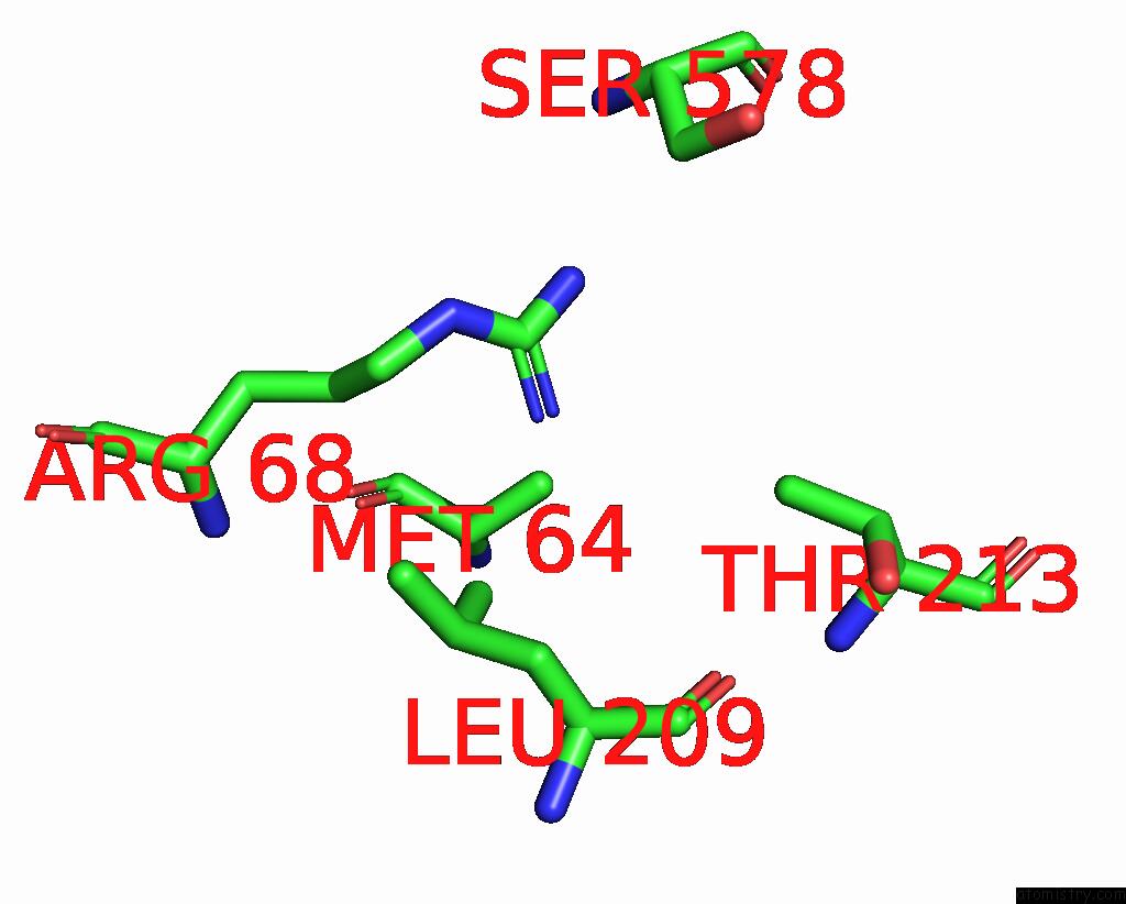
Platinum binding site 1 out of 2 in 8auc
Go back to
Platinum binding site 1 out
of 2 in the Structure of Peptidoglycan Hydrolase CG1735 From Corynebacterium Glutamicum, Trigonal Crystal Form
Mono view
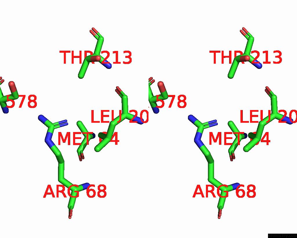
Stereo pair view
Mono view
Stereo pair view
A full contact list of Platinum with other atoms in the Pt binding
site number 1 of Structure of Peptidoglycan Hydrolase CG1735 From Corynebacterium Glutamicum, Trigonal Crystal Form within 5.0Å range:
|
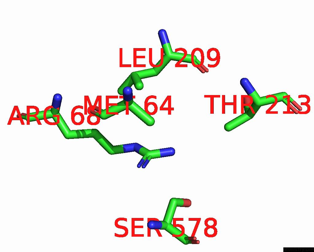
Platinum binding site 2 out of 2 in 8auc
Go back to
Platinum binding site 2 out
of 2 in the Structure of Peptidoglycan Hydrolase CG1735 From Corynebacterium Glutamicum, Trigonal Crystal Form
Mono view
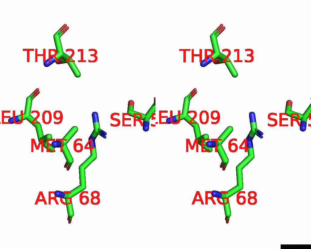
Stereo pair view
Mono view
Stereo pair view
A full contact list of Platinum with other atoms in the Pt binding
site number 2 of Structure of Peptidoglycan Hydrolase CG1735 From Corynebacterium Glutamicum, Trigonal Crystal Form within 5.0Å range:
|
Reference:
Q.Gaday,
D.Megrian,
G.Carloni,
M.Martinez,
B.Sokolova,
M.Ben Assaya,
P.Legrand,
S.Brule,
A.Haouz,
A.M.Wehenkel,
P.M.Alzari.
Ftsex-Independent Control of Ripa-Mediated Cell Separation in Corynebacteriales. Proc.Natl.Acad.Sci.Usa V. 119 99119 2022.
ISSN: ESSN 1091-6490
PubMed: 36469781
DOI: 10.1073/PNAS.2214599119
Page generated: Thu Oct 10 11:51:35 2024
ISSN: ESSN 1091-6490
PubMed: 36469781
DOI: 10.1073/PNAS.2214599119
Last articles
Fe in 2YXOFe in 2YRS
Fe in 2YXC
Fe in 2YNM
Fe in 2YVJ
Fe in 2YP1
Fe in 2YU2
Fe in 2YU1
Fe in 2YQB
Fe in 2YOO